Showing posts with label Hinglish. Show all posts

Diabetic Nephropathy Part - 1 (Hinglish)

Diabetic Nephropathy: Ek Comprehensive Guide


Introduction 

Diabetic nephropathy, ko diabetic kidney disease (DKD) ke naam se bhi jaana jaata hai, ye ek serious complication hai jo diabetes se affected logon ko hota hai. Ismein kidney damage ho jati hai jo lambe samay tak blood sugar ko theek se control na karne ke wajah se hota hai. Yeh condition kidney ke function ko affect karti hai, aur yeh aage chalke chronic kidney disease (CKD) ya end-stage renal disease (ESRD) tak le ja sakti hai. Basically, diabetic nephropathy ka matlab hai kidney function ka dheere-dheere khatam hona, jo high blood sugar ke harmful effects ki wajah se hota hai.


Historical Background  

Diabetes aur kidney disease ke beech ka connection pehli baar 18th century ke end mein dekha gaya jab diabetes ke patients ke urine mein protein (proteinuria) paya gaya. 1930s tak, Kimmelstiel aur Wilson ne diabetes aur kidney damage ke link ko aur clarify kiya, aur diabetic patients mein proteinuria aur hypertension ke saath nodular glomerulosclerosis ke classic lesions ko describe kiya. Samay ke saath diabetic nephropathy ke mechanisms ko samajhne mein advancements hui hain, jaise hyperglycemia-induced inflammation aur oxidative stress. Aaj diabetic nephropathy ko duniya ke kai parts mein CKD aur ESRD ka leading cause maana jaata hai.


ETIOLOGY (Karan) 

Diabetic nephropathy ka exact cause abhi tak clear nahi hai, lekin kai factors hai jo is condition ko trigger karte hain. High blood sugar (hyperglycemia) ko kidney damage ka main culprit mana jaata hai, jo hyperfiltration, advanced glycation end-products, aur inflammatory cytokines ke activation ke zariye hota hai. Kai researchers diabetes ko ek autoimmune condition bhi samajhne lage hain, jisme immune system ka role, jaise toll-like receptors aur regulatory T-cells, highlight kiya gaya hai, especially type 1 aur type 2 diabetes mein.


Blood sugar ko control karna food intake, liver ke glucose production, aur body ka glucose ko use karne ya store karne ke process par depend karta hai. Insulin, jo pancreas banata hai, is process ko control karta hai. Jab insulin resistance hoti hai, toh body ko blood sugar control karne mein problem hoti hai, jisse hyperglycemia (high blood sugar) aur hyperlipidemia (high fat levels in blood) hota hai. Yeh pancreas ke insulin-producing β-cells ko stress mein daal deti hai, jisse yeh damage ho jaate hain.


High blood sugar, fatty acids, aur inflammation milke immune system ko activate karte hain, jo β-cells ko aur damage karta hai. Isse insulin resistance aur inflammation ka ek vicious cycle create hota hai, jo diabetic complications, including kidney damage, ko aur badhata hai. High blood sugar kuch proteins jaise transforming growth factor-beta (TGF-β) aur vascular endothelial growth factor (VEGF) ka release trigger karta hai, jo kidneys mein cellular changes aur scarring ko lead karta hai.


Genetics ka bhi role ho sakta hai diabetic nephropathy mein, jaise kuch ethnic groups, African Americans, Hispanics, aur American Indians mein kidney disease ka risk zyada hota hai diabetes ke chalte. Researchers maante hain ki yeh genetic factors ka result ho sakta hai, jise "thrifty genotype" hypothesis kaha gaya hai.


Recent research yeh bhi suggest karta hai ki folic acid ka lower level diabetic nephropathy ka risk badha sakta hai. Ek study mein yeh paaya gaya ki folic acid ka level low hone se diabetes walon mein kidney disease ka chance 20% tak badh jaata hai. Halanki, genetics aur environmental factors diabetic nephropathy mein clearly role play karte hain, lekin iske causes ko puri tarah samajhne ke liye aur research ki zarurat hai.


RISK FACTORS


1. Long-standing Diabetes (Type 1 aur Type 2) 

Diabetes jitne zyada time tak rahe, diabetic nephropathy ka risk utna hi zyada hota hai. Jo log 10-20 saal ya usse zyada samay se diabetes ke saath jee rahe hain, unme ye complication hone ka chance zyada hota hai, especially agar unka blood sugar poorly controlled raha ho.

2. Poor Glycemic Control 

Chronic hyperglycemia (high blood sugar) diabetic nephropathy ka primary driver hai. Lambe samay tak elevated blood sugar levels kidneys ke filtering units ko damage karte hain, jisse proteins urine mein leak hone lagte hain. Blood sugar ko theek se control mein rakhna nephropathy ke risk ko kam karne ke liye zaroori hai.

3. Hypertension  

High blood pressure ek aur bada risk factor hai diabetic nephropathy ke liye. Yeh kidneys ke blood vessels ko damage karta hai, jisse kidneys waste filter karne mein kamzor pad jaati hain aur kidney disease aur tez badhne lagta hai. Blood pressure ko control karna nephropathy ke advancement ko slow karne ke liye bahut zaroori hai.

4. Genetic Predisposition 

Kuch log genetic reasons ke wajah se diabetic nephropathy ke liye zyada prone hote hain. Studies ne yeh dikhaya hai ki family history of kidney disease hone se nephropathy hone ka chance badh jaata hai un logon mein jo diabetes ke patient hain. Genetic factors kaafi hath tak yeh decide karte hain ki kidneys high blood sugar aur blood pressure ka kaise response dete hain, jo kidney damage ke development aur progression mein madad karta hai.


Pathophysiology (Disease Kaise Develop Hoti Hai) 


Diabetic nephropathy kidneys ke filtering units, jise glomeruli kehte hain, mein teen bade changes laata hai. Sabse pehle, high blood sugar levels mesangial cells ko expand karne lagte hain, jo protein production mein badhoti ya un proteins mein changes ke wajah se hota hai. Doosra, glomeruli ko surround karne wali membrane thick hone lagti hai. Aur teesra, glomerular scarring hoti hai jisse glomeruli ke andar ka pressure badh jaata hai, jo kuch blood vessels ke widening ya doosron ke damage hone se hota hai. Yeh sab changes disease ke progress se connected hote hain.


Diabetic kidney disease ka main issue extracellular matrix ka buildup hota hai. Shuruaat mein yeh membrane ko thicken karta hai aur mesangium ko expand karta hai. Microscope ke neeche glomeruli mein solid material ka increase aur kabhi-kabhi bade circular structures, jise Kimmelstiel-Wilson nodules kehte hain, dekhne ko milte hain. Membranes mein albumin jaise protein deposits bhi milte hain, lekin yeh immune system involvement ka sign nahi hota. Advanced cases mein aur zyada thickening dekhne ko milti hai glomerular membranes ki.


Dusri kidney diseases ke comparison mein, diabetic nephropathy usually kidneys ko wahi size mein rakhti hai ya kabhi-kabhi badhata bhi hai. Lekin patients ko high blood pressure develop ho jata hai jo disease ko aur badhata hai. Obesity aur metabolic syndrome, jo diabetes mein common hain, blood pressure badhane mein aur kidney damage ka risk badhane mein contribute karte hain.


Studies ne yeh dikhaya hai ki jo log higher systolic blood pressure rakhte hain, especially jab ghar par measure kiya jaata hai, unme diabetic nephropathy ka risk zyada hota hai. Yeh risk younger diabetes patients mein aur zyada hota hai. Obesity sodium reabsorption ko badhake kidneys mein blood pressure ko raise karta hai, jisse kidney damage hoti hai. Time ke saath, yeh urine mein protein aur kidney function ke decline ko lead karta hai, jo untreated hone par kidney failure tak pahucha sakta hai.


Relationship Between Diabetes and Kidney Damage 

Kidneys body se waste aur extra fluid ko nikalne ka kaam karti hain. Diabetic nephropathy mein, prolonged high blood sugar levels kidneys ke small blood vessels ko damage karte hain, specifically glomeruli ko, jo filtration ke liye responsible hote hain. Is damage ke chalte kidneys ka filtration function weak ho jata hai, jisse proteins jaise albumin urine mein leak hone lagte hain.


Role of Proteinuria and Hypertension in Diabetic Nephropathy Progression 

Proteinuria, yaani urine mein excess protein ka hona, diabetic nephropathy ka hallmark sign hai, jo kidney function ke deterioration ko dikhata hai. Shuruaat mein, thode amount mein albumin urine mein detect ho sakta hai, is condition ko microalbuminuria kehte hain. Jaise-jaise disease progress karti hai, zyada amount mein protein urine mein leak hone lagta hai, jo overt proteinuria tak le jaata hai. Proteinuria kidney damage ko signal karta hai, lekin yeh inflammation aur kidney tissues ke scarring ko bhi badhata hai.

Hypertension (high blood pressure) diabetic nephropathy ka ek cause bhi hai aur uska result bhi. Yeh kidney function ke decline ko accelerate karta hai, kyunki yeh pehle se hi damaged blood vessels par aur pressure dalta hai kidneys ke andar. Proteinuria aur hypertension ka combination ek vicious cycle create karta hai, jisme ek doosre ko aur badhate hain, kidney damage ko aur tez karte hue CKD and ESRD cause karte hain.


ARCANOBACTERIUM HAEMOLYTICUM INFECTION (Hinglish)

ARCANOBACTERIUM HAEMOLYTICUM INFECTION

Arcanobacterium haemolyticum ek bacterium hai jo 1946 mein pehli baar identify kiya gaya tha. Yeh primarily pharyngitis aur skin rashes jaise infections ka kaaran banta hai, aur khaaskar US military personnel aur South Pacific ke indigenous communities mein paya gaya hai. Shuruaat mein ise Corynebacterium pyogenes ka subspecies mana gaya tha, lekin baad mein iske distinct characteristics ke kaaran ise nayi genus Arcanobacterium mein reclassify kiya gaya, jiska matlab hai "Is bacterium ko 'chhupa hua bacteria' kaha gaya. Vaqt ke sath, ise sepsis aur osteomyelitis jaise gambhir infections se bhi sambandhit kiya gaya hai."


SYMPTOMS AUR PRESENTATION

A. haemolyticum infection ka sabse common symptom sore throat hai, jo lagbhag sabhi patients (97-100%) mein report kiya jata hai. Dusre symptoms mein shamil hain:

  • Itchy skin (33%)
  • Nonproductive cough (33%)
  • Fever
  • Rash

Yeh rashes arms aur legs se shuru hote hain, lekin chehre, palms aur soles ko nahi chhute. Rash 48 ghante tak reh sakta hai, aur recovery ke dauran halki peeling dekhne ko milti hai. Ek review mein, 50% se zyada patients mein tonsillar exudates, fever, aur skin rash dekha gaya.


TRANSMISSION AUR RISK FACTORS

Ab tak koi confirmed risk factors nahi mile hain, lekin yeh speculate kiya gaya hai ki cows aur buffaloes jaise animals ke contact mein aane ya unpasteurized milk ke consumption se infection ka risk badh sakta hai. Bacterium European bison mein bhi paya gaya hai, lekin transmission ka tareeka ab tak unclear hai. Human-to-human transmission ka bhi shak hai, lekin proper hygiene, jaise handwashing, se iske spread ko roka ja sakta hai.


COMPLICATIONS

A. haemolyticum infection ke complications mein peritonsillar abscesses, sepsis, aur skin aur soft tissue infections shamil ho sakte hain. Severe sore throat aur fever ke cases mein hospitalization ki zarurat pad sakti hai jisme abscess drainage ya intravenous antibiotics ki madad li jaati hai.


DIAGNOSIS

A. haemolyticum ka diagnosis mushkil ho sakta hai, kyunki iski colonies slow-growing hoti hain aur routine throat cultures mein miss ho sakti hain. Specialized blood agar aur prolonged incubation (48 ghante tak) ki zarurat padti hai isse detect karne ke liye. Kuch cases mein, rashes se li gayi skin biopsies mein inflammation dikhayi deti hai, lekin bacteria khud nahi milta.


ARCANOBACTERIUM HAEMOLYTICUM INFECTION KA TREATMENT

A. haemolyticum ek significant lekin underrecognized bacterium hai jo mild pharyngitis se lekar severe invasive diseases, jaise sepsis ya osteomyelitis tak infections ka kaaran ban sakta hai. Jaldi pehchan aur proper treatment complications ka risk kam karne mein madad karte hain. Niche A. haemolyticum infections ke treatment ke bare mein comprehensive guide di gayi hai.


1. Pharyngitis aur Mild Infections

A. haemolyticum ke zyada infections pharyngitis ke roop mein hote hain, jisme tonsillitis, fever, aur kabhi-kabhi rash bhi dikhai dete hain. Halke cases khud theek ho sakte hain, lekin antibiotic treatment symptoms ki duration kam karne aur complications se bachne ke liye recommend kiya jata hai.

Antibiotic Therapy:

  • Penicillin: Penicillin aur related antibiotics jaise amoxicillin standard treatments hote hain, lekin kuch cases mein bacteria ke intracellular survive karne ke kaaran resistance dekhne ko milta hai.
  • Macrolides (e.g., erythromycin, azithromycin): Macrolides ko intracellular bacteria ke against effective mana jata hai, aur yeh A. haemolyticum infections ke treatment ke liye first choice hote hain. Azithromycin ko iski convenient dosing schedule ke liye khaaskar pasand kiya jata hai.
  • Clindamycin: Penicillin allergy wale patients ya severe infections wale patients ke liye ek viable alternative hai. Clindamycin achhi tissue penetration ke saath anaerobic bacteria ke against effective hota hai.

Dosage Recommendations:

  • Tab. Erythromycin: 250-500 mg har 6-12 ghante ke liye, 7-10 din tak.
  • Tab. Azithromycin: Pehle din 500 mg, uske baad 4 din tak 250 mg roz.
  • Tab. Clindamycin: 150-450 mg har 6-8 ghante, infection ki severity ke hisaab se.

Expected Outcomes: Antibiotic therapy se pharyngitis usually 3 din mein theek ho jata hai. Bina treatment ke symptoms 2 hafte tak chal sakte hain.


2. Severe ya Systemic Infections

Zyada severe cases, jaise sepsis ya deep tissue infections, mein jaldi intravenous antibiotics ka intervention zaroori hota hai.

Intravenous Antibiotics:

Beta-lactams (e.g., penicillin, cefuroxime, cefotaxime): Yeh antibiotics severe cases jaise sepsis ya osteomyelitis ke liye first-line treatments hain. Yeh bacterial cell wall ke construction mein interfere karke bacteria ko kamzor karte hain aur uske growth ko rok dete hain.

Vancomycin: Severe infections mein beta-lactams allergy wale patients ya resistance ke shak par use hota hai. Lekin vancomycin ke resistance ke cases bhi report kiye gaye hain, isliye sensitivity testing zaroori hai.

Antibiotic Sensitivity: A. haemolyticum typically sensitive hota hai:

  • Minocycline
  • Vancomycin
  • Beta-lactams

Antibiotic Resistance: Resistance dekhne ko mili hai:

  • Gentamicin
  • Fluoroquinolones (e.g., levofloxacin, ciprofloxacin)
  • Trimethoprim-sulfamethoxazole
  • Tetracyclines (30% strains mein resistance)

Treatment Duration: Severe infections ke liye intravenous therapy usually 2-6 hafton tak chalti hai, infection ki site aur severity ke hisaab se.


3. Complications ka Management

Kuch cases mein, untreated ya severe infections peritonsillar abscesses ya sepsis jaisi complications tak pahunch sakti hain.

  • Peritonsillar Abscess: Is condition mein surgical drainage ki zarurat padti hai, jiske baad antibiotics (penicillin, clindamycin, ya cefuroxime) 7-14 din tak diye jate hain.
  • Sepsis: Immediate intravenous antibiotics aur supportive care zaroori hota hai. Blood cultures sensitivity results ke hisaab se appropriate antibiotics select karne mein madad karte hain.


4. Adjunct Therapies

Supportive treatments antibiotic therapy ko complement kar sakti hain, khaaskar pharyngitis ya systemic infections ke cases mein.

  • Antipyretics aur Analgesics: Non-steroidal anti-inflammatory drugs (NSAIDs) jaise ibuprofen ya acetaminophen fever aur pain manage karne mein madad karte hain.
  • Antiseptic Lozenges: Amylmetacresol jaise antiseptics wale lozenges bacterial load kam karne aur symptoms ko soothe karne mein madad karte hain.


5. Hospital Admission aur Parenteral Therapy

  • Systemic ya severe infections wale patients ko hospitalization ki zarurat ho sakti hai, khaaskar agar wo swallow nahi kar pa rahe ho ya sepsis jaise complications ka risk ho.
  • Intravenous Antibiotics: Aise patients ko parenteral antibiotics ki zarurat hoti hai aur infection resolve hone tak close monitoring ki jati hai.


Monitoring aur Follow-Up

Patients ko antibiotics shuru karne ke 48-72 ghante ke andar reassess kiya jana chahiye taaki improvement ensure kiya ja sake. Severe cases mein, repeat blood cultures aur imaging required ho sakti hai recovery confirm karne ke liye.


PREVENTION

A. haemolyticum ke liye koi specific preventive measures recommended nahi hain, lekin achhi hygiene maintain karna, shared utensils avoid karna, aur proper food handling (khaaskar dairy products ke saath) infection transmission ke risk ko kam kar sakte hain.


CONCLUSION 

Early detection aur appropriate treatment critical hote hain Arcanobacterium haemolyticum infections ka sahi management aur complications ko prevent karne ke liye.

Black Lung Disease (Hinglish)

Black Lung Disease (Coal Workers’ Pneumoconiosis)


Introduction

Black Lung Disease, jise Coal Workers’ Pneumoconiosis (CWP) bhi kaha jata hai, ek chronic lung disease hai jo coal dust ke lambi samay tak inhale karne se hoti hai. Ye sthiti mukhya roop se coal miners ko prabhavit karti hai aur lungs mein dust ka accumulation kar deti hai, jo inflammation, scarring, aur severe cases mein lung function ko impair karti hai. "Black lung" shabd un lungs ke dark appearance se aata hai jo post-mortem coal dust deposits ki wajah se hota hai.


Chest X ray of diagnosed silicosis patient


Causes (Karan)

Black Lung Disease ka mukhya karan coal dust ke lambi avadhi tak ka exposure hai. Lekin kuch factors kisi vyakti ke risk ko badha sakte hain:

  • Coal dust ka lamba exposure: Jitna jyada dust samay ke saath inhale kiya jata hai, utna hi risk badh jata hai.
  • Protective gear ki kami: Respiratory protection ka na hona ya uska galat istemal disease develop hone ke chances ko badhata hai.
  • Poor ventilation: Mining environments jahan ventilation thik nahi hota, wahaan coal dust ke concentration jyada ho jate hain, jo risk ko badha dete hain.


CT of silicosis patient


Black Lung Disease ke types and stages 

Simple CWP: 

Iske prarambhik charan mein, disease lungs mein choti nodules bana deta hai. Kai logon ko simple CWP hone par symptoms nahi dikhte, lekin exposure jaari rehne par disease aage badh sakti hai. 

Complicated CWP ya Progressive Massive Fibrosis (PMF): 

Ye advanced stage hai jisme lungs mein large masses of scar tissue (fibrosis) bante hain, jo lung function ko severely limit karte hain aur jisse persistent coughing, breathlessness, aur potential respiratory failure jaisi symptoms hoti hain.


Symptoms 

Black Lung Disease ke symptoms stage ke aadhar par alag hote hain:

Prarambhik Symptoms: Physical exertion ke dauran persistent coughing aur shortness of breath. 

Advanced Symptoms: 

  • Chronic cough, kabhi kabhi dark-colored sputum ke sath.
  • Constant shortness of breath, jabki resting ke dauran bhi.
  • Chest pain, fatigue, aur daily tasks karne mein dikkat respiratory limitations ki wajah se.
  • Severe cases mein, respiratory failure.


Diagnosis 

Diagnosis mein multiple methods ka istemal hota hai lung damage ke extent ko assess karne ke liye:

  1. Chest X-rays: Ye nodules aur fibrosis ke signs ko detect karne ke liye istemal hoti hain.
  2. Pulmonary function tests: Ye lung capacity aur airflow ko measure karte hain taaki impairment ka degree pata chale.
  3. CT scans: Ye lung scarring ki severity ko assess karne ke liye detailed imaging dete hain.
  4. Medical history: Doctors patient ki coal dust exposure, smoking habits, aur dusre occupational risks ka evaluation karte hain.


Black Lung Disease ke Treatment Options 

Black Lung Disease ka koi cure nahi hai, lekin symptoms ko manage karna aur further damage se bachna bahut zaroori hai: 

1. Medications for Black Lung Disease: 

Bronchodilators: Ye medications airways ke around muscles ko relax karne mein madad karte hain, jisse woh khul jaate hain aur patients ko saans lene mein aasan hoti hai. Ye symptoms jaise shortness of breath aur chest tightness ko kam karne mein madadgar hote hain. Kuch commonly used bronchodilators hain:

  • Albuterol (Proventil, Ventolin): Ye ek fast-acting bronchodilator hai jo breathing difficulties ke quick relief ke liye inhalers mein istemal hota hai.
  • Salmeterol (Serevent): Ye ek long-acting bronchodilator hai jo extended periods tak airways ko khula rakhne mein madad karta hai, isse symptoms ko control karne ke liye regular istemal karna hota hai. 
  • Tiotropium (Spiriva): Ye bhi ek long-acting bronchodilator hai jo airways ko khula rakhne mein aur breathing difficulties ko long term mein reduce karne mein madad karta hai.

Ye medications aam taur par inhalers ya nebulizers ke madhyam se directly lungs tak pahunchaya jata hai aur fast symptom relief dete hain. 

2. Oxygen therapy: 

Jaise jaise lung function kharab hota hai, kuch patients ko blood oxygen levels ko maintain karne ke liye supplemental oxygen ki zaroorat pad sakti hai. Ye breathlessness aur fatigue ke symptoms ko relieve karne mein madad karta hai. 

3. Pulmonary rehabilitation: 

Is program mein exercises, breathing techniques, aur education shamil hoti hai taaki patients apne symptoms ko manage kar sakein aur lung capacity ko improve kar sakein. 

4. Avoidance of further exposure: 

Sabse important step ye hai ki coal dust ke further exposure se bacha jaye, jo disease ke worsen hone se rok sakta hai. Mining work se retire hona aur similar environments mein respiratory protection ka istemal karna zaroori hai.


Complications 

  1. Chronic Obstructive Pulmonary Disease (COPD): Kai logon ko Black Lung Disease hone par COPD develop hota hai, jo airflow aur lung function ko aur limit karta hai.
  2. Heart problems: Black Lung Disease ki wajah se lungs mein badh gaye pressure se pulmonary hypertension hota hai, jo heart strain aur potential heart failure ki taraf le ja sakta hai.
  3. Increased mortality: Disease ke advanced stages respiratory failure ki taraf le ja sakte hain, jo life expectancy ko significantly reduce karte hain.


Prevention 

Black Lung Disease se bachne ka sabse achha tareeqa coal dust exposure ko kam karna hai:

  • Dust control in mines: Coal dust levels par regulations ko lagu karna aur enforce karna bahut zaroori hai Black Lung Disease ko prevent karne ke liye. Dust suppression techniques ko har jagah istemal karna chahiye.
  • Proper protective gear: Miners ko consistently respiratory protection ka istemal karna chahiye taaki inhale hone wale dust ki matra kam ho sake.
  • Health surveillance: Regular medical check-ups, including lung function tests, early signs of Black Lung Disease ko detect karne ke liye madadgar hote hain, jisse early intervention possible hota hai.


Prognosis 

Black Lung Disease se prabhavit vyaktiyon ka prognosis is baat par depend karta hai ki disease kis stage par diagnose kiya gaya hai aur kya coal dust ke further exposure ko roka gaya hai. Early intervention aur consistent management se quality of life improve ki ja sakti hai, lekin ye disease progressive hai, aur advanced stages significant respiratory aur heart issues ki taraf le ja sakti hain, jo life expectancy ko ultimately reduce karte hain.


Conclusion (Nishkarsh) 

Black Lung Disease coal mining industry mein ek serious occupational health issue hai. Jabki treatment symptoms ko manage karne aur disease ke progression ko slow karne mein madadgar ho sakta hai, prevention dust control aur proper protective measures ke through key hai. Stronger health aur safety regulations ke liye ongoing advocacy, saath hi affected workers ke liye legal support, is preventable lekin incurable disease se nipatne ke liye zaroori hai.

National Mental Health Programme (Hinglish)

National Mental Health Programme (NMHP)


Introduction 

Mental health issues duniya bhar mein kaafi aam hain aur yeh har tarah ke logon ko prabhavit karti hain. Bahut se adults kabhi na kabhi anxiety, fatigue, ya sleeplessness jaise lakshan anubhav karte hain, aur har saat mein se ek vyakti ko apne jeevan mein kisi na kisi roop mein ek diagnosable mental health disorder ka samna karna padta hai.


Burden of Disease 

1993 mein World Bank ke ek report ne bataya ki mental health disorders, healthy life years ke loss ka karan bante hain, aur yeh diarrhea, malaria, worm infestations aur tuberculosis se zyada khatarnak hain jab inhe alag-alag dekha jaye. 2020 tak, yeh anuman lagaya gaya tha ki mental health conditions global burden of disease ka 15% hissa hongi.

Bharat mein pichle 20 saalon mein kiye gaye kai epidemiological studies yeh dikhate hain ki psychiatric disorders yahan utne hi aam hain jitne videsh mein. In disorders ki prevalence 18 se 207 per 1,000 logon ke beech hai, aur iska average 65.4 per 1,000 hai. Kisi bhi samay, lagbhag 2-3% jansankhya gambhir aur asamarthak mental health conditions ya epilepsy se joojh rahi hoti hai. Durbhagya se, inmein se adhiktar log rural areas mein rehte hain jahan mental health care ki facilities ki kami hai. Saath hi, general outpatient departments (OPD) mein aaye hue kai adults ko mental health issues ka samna hota hai, lekin kai cases miss ho jate hain kyunki health care providers aksar mental health ke symptoms ke baare mein sawal nahi poochte, jiska nateeja unnecessary tests aur treatments hota hai.


The National Mental Health Programme 

Mental health ka significant burden aur sevaon ki kami ko dekhte hue, Bharat sarkar ne 1982 mein National Mental Health Programme (NMHP) launch kiya.

Is programme ka dhyan teen mukhya kshetron par hai:

1. Treatment un logon ke liye jo mental illness se grasit hain.

2. Rehabilitation unke liye jo prabhavit hue hain.

3. Positive mental health ke liye prevention aur promotion.


Aims 

NMHP ke aims hain:

  • Mental aur neurological disorders ke saath unse judi disabilities ka prevention aur treatment karna.
  • Mental health care ko general health services mein integrate karna mental health technology ke madhyam se.
  • National development mein mental health ke siddhanton ka istemal karke quality of life ko behtar banana.


Objectives 

NMHP ke pramukh objectives hain:

  • Sabhi logon ke liye, khaaskar kamzor aur vanchit varg ke logon ke liye, minimum mental health care services ki uplabdhata aur pahuch sunishchit karna.
  • General health care aur social development mein mental health gyaan ka prayog ko badhava dena.
  • Community ko mental health services ke vikas mein bhaagidari ke liye prerit karna aur self-help initiatives ko badhava dena.


Strategies 

In aims ko paane ke liye programme kuch mukhya strategies ka palan karta hai:

  • NMHP ke madhyam se mental health care ko primary health care ke saath integrate karna.
  • Mental disorders ke treatment ke liye tertiary care institutions ka pravadhan karna.
  • Mental illness ke stigma ko mitane aur mental health conditions wale logo ke adhikaron ka rakshan karne ke liye Central Mental Health Authority aur State Mental Health Authorities jaise sansthano ka gathan karna.


Mental Health Care 

Mental health treatment ko healthcare system ke vibhin staron par prathmikta di gayi hai, jaise ki:

  1. Gaon aur sub-center level par primary care
  2. Primary Health Centers (PHC)
  3. District hospitals
  4. Mental hospitals aur psychiatric teaching units


District Mental Health Programme 

Yeh programme district level par mental health care par kendrit hai, jisme nimnalikhit components shamil hain:

  • Har state mein identified Nodal Institutes par mental health team ke sabhi karyakartao ka training programme.
  • Mental health ke prati jagrukta badhane aur stigma kam karne ke liye public education campaigns.
  • Jaldi diagnosis aur treatment ke liye OPD aur indoor services ka pravadhan.
  • Community se mila data aur anubhav, state aur central governments ko future planning, services mein sudhar aur research ke liye pradan karna.


Support and Funding 

World Bank aur WHO jaise antar-rashtriya sansthano ko programme ke vibhin components mein support dene ke liye sampark kiya gaya hai. Bharat sarkar dwara state governments aur nodal institutes ko pehle paanch saal tak funding pradan ki jati hai, jisme staff ki tankhwa, equipment, gaadi, dawa, stationary, training aadi ka kharcha shamil hota hai. Paanch saal ke baad, in sansthano ko apne aap chalana hota hai. Central Mental Health Authority, jo Bharat sarkar dwara gathit hai, Mental Health Act 1986 ke implementation ko monitor karti hai. State Mental Health Authorities ko bhi aise hi karya karne ke liye gathit kiya gaya hai.


Monitoring and Oversight 

National Human Rights Commission bhi Bharat sarkar ke saath milkar mental hospitals mein conditions ko monitor karta hai, taki quality care ko sunishchit kiya ja sake aur joint studies ke recommendations ke anusaar sudhar kiya ja sake.


Hand, Foot, and Mouth Disease in Children (HFMD)


Hand, Foot, and Mouth Disease in Children


Introduction 

Hand, foot, aur mouth disease (HFMD) ek common viral bimari hai jo mainly chhote bachchon ko affect karti hai. Yeh usually mild hoti hai, lekin kabhi-kabhi discomfort aur complications ka kaaran ban sakti hai. HFMD viruses ke enterovirus family ke karan hoti hai, jisme se coxsackievirus A16 aur enterovirus 71 sabse common hain.


Symptoms 

Virus ke exposure ke 3 se 7 din baad symptoms nazar aane lagte hain, jo include karte hain:

1. Fever: Pehla sign hota hai, jo usually mild se moderate hota hai.

2. Sore Throat: Gala mein dard ho sakta hai, aur nighalne mein bhi takleef ho sakti hai.

3. Mouth Sores: Dardnakh zakhm munh mein bante hain, jo zyadaatar jeebh, masoodon, aur andar ke gaalon par hote hain.

4. Rash: Haathon aur paon par ek rash bhi dikhayi de sakta hai. Yeh daag usually laal hote hain aur blister ban sakte hain.

5. Loss of Appetite: Munh ke sores ke wajah se bachche khana ya pani nhin lete hain.

6. Fatigue aur Irritability: General thakaan aur chidchidaapan hota hai, jo chhote bachchon mein zyada dikhai deta hai.


Transmission 

HFMD chhune se phailne waali bimari hai, aur yeh is tarah se spread hoti hai:

  • Direct Contact: Infected surfaces ya fluids (jaise saliva, blister ka fluid) ko touch karna.
  • Respiratory Droplets: Kisi bimaar shakhs ke khansi ya cheekhne se.
  • Feco-Oral Route: Virus stool mein bhi hota hai, to safai ki kami se yeh phail sakta hai.


Diagnosis 

Diagnosis usually in baaton par depend karta hai:

  • Clinical Evaluation: Doctor symptoms ko assess karte hain aur characteristic mouth sores aur rashes dekhte hain.
  • Recent Illness History: Schools ya daycare centers mein recent outbreaks ko note karna helpful ho sakta hai.
  • Kabhi-kabhi, laboratory tests ki zarurat ho sakti hai taaki virus ka type confirm ho sake.


Bacchon mein Hand, Foot, aur Mouth Disease (HFMD) ka ilaj 

HFMD ka koi specific antiviral treatment nahi hai, par symptoms ko manage karne aur supportive care dene par focus kiya jata hai. Yahan kuch key treatment strategies di gayi hain:

1. Symptom Relief 

Pain Relief - Over-the-Counter Medications:

  • Acetaminophen (Tylenol): Bukhar kam karne aur dard se rahat dene ke liye.
  • Ibuprofen (Advil, Motrin): Bukhar aur discomfort kam karne ka ek aur option.
  • Note: Bacchon ko aspirin dena avoid karein kyunki Reye's syndrome ka risk hota hai.

Mouth Sores:

  • Topical Anesthetics: Benzocaine wale gels ya liquids temporary dard ko numb kar sakte hain. Yeh sores par directly lagaye ja sakte hain.
  • Saltwater Rinses: Namak aur garam paani ka solution gala aur munh ke sores ko soothe karne mein madad karta hai (lekin chhote bachchon ke liye use karna mushkil ho sakta hai).

2. Hydration 

  • Fluid Intake Encourage Karein: Bacchon ko hydrated rakhna bahut zaruri hai, khaaskar jab unhe munh ke sores ke kaaran nighalne mein mushkil hoti hai.
  • Thande Fluids Offer Karein: Ice chips, popsicles, ya smoothies soothing ho sakte hain aur hydration mein madad karte hain.
  • Acidic ya Spicy Foods/Drinks Avoid Karein: Yeh munh ke sores ko irritate kar sakte hain.

3. Nutrition 

Soft Foods Encourage Karein: Aise foods khilayein jo soft aur easily nighalne wale ho, jaise:

  • Mashed potatoes
  • Applesauce
  • Yogurt
  • Oatmeal 
  • Hard ya Crunchy Foods Avoid Karein: Aise foods jo munh ke sores ko irritate kar sakte hain, avoid karein.

4. Fever Management

Temperature Monitor Karein: Bacchon ka temperature track karein aur zarurat padne par fever-reducing medications de.

Comfort Measures: Bacchon ko lightweight kapde pehnayein aur unke aas-paas ka environment thanda rakhein taaki fever ko manage kiya ja sake.

5. Rest aur Comfort

Rest Encourage Karein: Bacchon ko rest karne ke liye ek quiet aur comfortable environment dein.

Distraction Techniques: Aise activities jaise reading, quiet games, ya movies dekhna use karein taaki bacchon ko recovery ke dauraan busy rakha ja sake.

6. Follow-Up Care 

Symptoms par nazar rakhein: Bacchon ki tabiyat ko gahraai se observe karein. Agar lakshan bigadne lagein, pani ki kami, gambhir symptoms ya phir uncha bukhar dikhai de, toh turant chikitsak se salaah lein.


Healthcare Provider se Salah Len: Agar dehydration ya severe symptoms ka darr ho, khaaskar chhote bacchon ya jinhe already health issues hain, unme doctor se consult karna zaruri hai.


Prevention 

Preventive measures mein include hota hai:

  • Achhi Safai ki Adatein: Bathroom use karne ke baad aur khane se pehle sabun se haath dhona.
  • Close Contact Avoid Karein: HFMD wale bacchon ko school ya daycare se door rakhein jab tak wo contagious na ho (usually jab fever theek ho jaye aur munh ke sores heal ho jaayein).
  • Surfaces Disinfect Karein: Toys aur frequently touched surfaces ko clean karke virus ke spread ko kam kiya ja sakta hai.


Complications 

HFMD usually mild hota hai, lekin kabhi-kabhi complications bhi ho sakte hain, jaise:

  • Dehydration: Munh ke sores ke kaaran nighalne mein dikkat ke karan.
  • Viral Meningitis: Kabhi-kabhi virus se serious neurological conditions ho sakti hain.
  • Myocarditis ya Encephalitis: Yeh serious complications rare hote hain, lekin enterovirus infections mein ho sakte hain.


Prognosis 

HFMD usually 7 se 10 din mein bina medical intervention ke theek ho jata hai. Zyadaatar bacche bina koi long-term health issues ke fully recover karte hain.


Conclusion 

Hand, foot, aur mouth disease ek common, generally mild viral infection hai bacchon mein. Symptoms, transmission, aur preventive measures ka dhyaan rakhke outbreaks ko effectively manage kiya ja sakta hai, aur illness ke dauraan bacchon ko comfortable rakha ja sakta hai. Agar severe symptoms ya dehydration ka sign dikhe, toh turant medical attention leni chahiye.


ACTINOMYCOSIS (Hinglish)

 ACTINOMYCOSIS


Introduction:   

Actinomycosis ek chronic bacterial infection hai jo 'Actinomyces' bacteria se hota hai. Ye bacteria filamental, gram-positive hote hain, jo aam taur pe mouth, digestive system, aur female genital tract mein paye jaate hain. Lekin jab mucous membrane damage hoti hai, jaise dental surgery, abdominal injury, ya IUD use karne ke baad, to infection shuru ho sakta hai.


Actinomycosis kaise develop hota hai:   

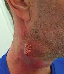
Ye infection dheere dheere tissues mein failta hai aur abscesses aur sinus tracts banata hai. Kabhi kabhi is infection ke discharge mein sulfur granules (chhoti yellow particles) bhi dekhe ja sakte hain. Ye infection alag-alag jagah affect kar sakta hai jaise face (cervicofacial), chest (thoracic), ya abdomen aur pelvis. Isko diagnose karna mushkil hota hai kyunki ye tumors ya tuberculosis jaisi bimariyon jaise lagta hai.


Common types:   

1.  Cervicofacial Actinomycosis (Lumpy Jaw): Ye sabse common type hai aur aksar poor dental hygiene ya oral surgery se judta hai. Jaw mein swelling hoti hai, fistulas ban jaate hain, aur pus discharge hota hai jisme sulfur granules hoti hain.

2.  Thoracic Actinomycosis: Ye lungs ko affect karta hai, aksar bacteria inhale karne ke baad. Chest pain, cough, aur kabhi kabhi chest wall pe sinus tracts bhi ban jate hain.

3.  Abdominal aur Pelvic Actinomycosis: Surgery ya abdominal injury ke baad hota hai ya phir women mein IUD use ke wajah se hota hai. Abdominal pain, bowel habits mein changes, aur mass ki tarah feel hota hai, jise galat tareeke se cancer samajh liya jaata hai.


Symptoms:

  1. Cervicofacial Actinomycosis:  Jaw mein swelling, skin discoloration, sinus discharge, aur chewing mein dikkat.
  2. Thoracic Actinomycosis:  Cough, chest pain, shortness of breath, weight loss, aur fever.
  3. Abdominal Actinomycosis:  Abdominal pain, nausea, weight loss, aur abdomen mein mass ka sensation.
  4. Pelvic Actinomycosis:  Lower abdominal pain, vaginal bleeding ya discharge.


Diagnosis:   

Iska diagnosis karna thoda challenging hota hai. Abscesses ya sinus tract se tissue samples lene padte hain aur sulfur granules discharge mein milte hain to infection ka confirmation hota hai. Imaging tests jaise CT scan mass-like infection dikhate hain jo cancer jaise lag sakte hain. Laboratory tests anemia, elevated inflammatory markers show karte hain aur 'Actinomyces' ko cultures mein detect kar sakte  hain.


Treatment for Actinomycosis:

Is infection ka treatment antibiotics ke zariye kiya jaata hai.  Penicillin G  sabse zyada effective hoti hai. Treatment mein high doses antibiotics ka istamaal kiya jaata hai jo 6 mahine se lekar 1 saal tak chal sakta hai. Severe cases mein antibiotics ke saath surgery bhi zaroori ho sakti hai.


Antibiotic Treatment:

1. Penicillin G (Pfizerpen, Bicillin): Severe cases mein pehle intravenous (IV) Penicillin G diya jaata hai. Phir, oral antibiotics jaise Penicillin VK pe switch kiya jaata hai jab tak treatment complete na ho. Treatment 6 se 12 mahine tak chal sakta hai, ye infection ki severity aur response pe depend karta hai.

2.  Alternative antibiotics: Agar kisi ko penicillin se allergy ho, to doosre antibiotics ka istamaal kiya jaata hai jaise:

  • Doxycycline:  Ye bacterial protein synthesis ko rokta hai. Penicillin-allergic patients ke liye use hota hai, lekin pregnant women ke liye avoid kiya jaata hai.
  • Clindamycin (Cleocin):  Penicillin-allergic patients ke liye doosra option. Ye bacterial protein synthesis block karta hai, lekin companion bacteria ko cover nahi kar pata.
  • Amoxicillin/Clavulanic acid (Augmentin):  Mild to moderately severe cases mein use hota hai.
  • Ceftriaxone (Rocephin):  Moderate to severe infections mein use hota hai, jaise cervicofacial aur thoracic actinomycosis.
  • Imipenem/Cilastatin (Primaxin):  Severe abdominal ya pelvic infections ke liye kaafi effective hota hai.


Surgical Treatment:   

Jahan antibiotics se kaam nahi hota, wahan surgery zaroori ho sakti hai. Surgery ke zariye bade abscesses drain kiye jaate hain, infected tissue remove kiya jaata hai, aur kuch complications jaise fibrotic lesions ko treat kiya jaata hai. Kabhi kabhi abdominal actinomycosis mein patients ki emergency surgery ki zaroorat hoti hai agar peritonitis ya bowel perforation ho jaaye.


Monitoring During Treatment:   

Kyuki treatment kaafi lambe time tak chal sakta hai, regular follow-up zaroori hota hai jaise CT scan ya MRI ke zariye infection ka response monitor karna. Blood tests bhi karaye jaate hain recovery aur side effects dekhne ke liye.


Prevention:   

Achhi oral hygiene aur regular dental care important hai, khaaskar oral surgery ke baad. Aur IUD use karne wali women ko pelvic actinomycosis ke risk ko dhyaan mein rakhna chahiye.


Complications:   

Agar untreated chhoda gaya, to actinomycosis bone infections, brain abscesses, aur endocarditis (heart infection) jaise serious complications tak le ja sakta hai. Lekin early symptoms ko pehchaankar aur prolonged antibiotics course follow karke zyada tar patients achhe se recover karte hain.


Acrodermatitis Chronica Atrophicans (Hinglish)


 Acrodermatitis Chronica Atrophicans (ACA)


Introduction:

Acrodermatitis chronica atrophicans (ACA) European Lyme borreliosis ka late ya third stage hai, jo 'Borrelia afzelii' ke infection se hota hai. Is condition ko pehli baar 1883 mein identify kiya gaya aur 1902 mein describe kiya gaya. ACA ek progressive skin condition hai jisme fibrosis aur tissue atrophy hoti hai, jismein tissue paper jaise lagte hain.


Pathophysiology aur Etiology of Acrodermatitis Chronica Atrophicans (ACA)

Causative Agents:

ACA ka primary cause Borrelia afzelii hota hai, lekin Borrelia garinii (Borrelia burgdorferi sensu lato complex ka dusra genospecies) bhi implicated hai. Ye spirochetes humans mein tick ke kaatne se transfer hote hain aur Lyme borreliosis ka cause bante hain.


aca
Borrelia afzelii


 Pathophysiology:

Lyme disease ke doosre forms ke opposite, ACA khud se thik nahi hoti, jo ek unique pathogenic mechanism ko dikhata hai. Iska exact process abhi tak puri tarah se samjha nahi gaya, lekin kuch factors Borrelia ke skin mein persist karne ke liye zimmedar hain:

1.  Spirochete Persistence:

Complement system ke resistance: Bacteria immune response se bache rehte hain.

Immune evasion: Spirochetes immunologically protected sites jaise endothelial cells aur fibroblasts mein chhupe rehte hain.

Antigenic variation: Pathogen apne surface antigens ko badal leta hai, jisse immune system effective response nahi de pata.

2.  Immune Dysfunction:

  • Inadequate antibody response: ACA patients ke antibodies ka spectrum narrow hota hai aur protective antibodies nahi bante.
  • MHC Class II molecules ka downregulation: Langerhans cells (jo skin ke immune cells hote hain) mein in molecules ki kami T-cells activation ko impair karti hai.
  • Cytokine expression: Restricted cytokine profile jaise interferon-gamma ka absence, immune system ko infection se effectively fight karne se rokta hai.

3.  Autoimmune Considerations:  Cross-reactive antibodies tissue damage mein yogdan karte hain, lekin autoimmunity ka role abhi clear nahi hai. Chronicity ek aise immune response se judi ho sakti hai jo inadvertently host tissues ko harm karta hai.


Atrophic Skin Changes ka Pathogenesis:

Exact mechanism jo ACA ke characteristic skin atrophy ka cause banta hai wo samjha nahi gaya hai, lekin yeh mana jata hai ki periarticular sites low skin temperatures ya oxygenation ki wajah se Borrelia spirochetes ke liye favorable environment provide karate hain.


Clinical Presentation:

ACA aksar extremities pe aati hai, khaas taur pe extensor surfaces par, aur yeh do distinct phases mein hoti hai:

1.  Inflammatory Phase:

  • Bluish-red skin discoloration aur swelling.
  • Soft, painless, poorly demarcated plaques jo milkar ek ho sakti hain.
  • Condition distal extremities se start hokar proximally spread hoti hai.


2.  Atrophic Phase:

  • Mahino ya saalon baad hoti hai, jisme dark red ya brown discoloration hoti hai.
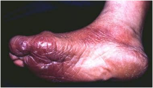
  • Skin thin, translucent, aur wrinkled ho jati hai, cigarette paper jaisi dikhai deti hai.
  • Prominent blood vessels, hair loss, aur sebaceous ya sweat glands ka absence hota hai.


Associated Symptoms:

Fibrotic nodules elbows, knees, aur adjacent joints pe form hote hain. Skin ulcerate kar sakti hai, aur malignant lesions develop ho sakte hain. Fingers aur toes mein deformities bhi ho sakti hain agar untreated chhod diya jaye.

Systemic Involvement:

Neurological involvement common hai, jisme sensory polyneuropathy symptoms jaise pain, paresthesia, aur paresis (partial paralysis) shamil hote hain.


Diagnosis:

Detailed clinical history, epidemiologic data, histopathology aur serologic testing ke through diagnosis kiya jata hai. ELISA, indirect immunofluorescence, aur Western blot assays commonly use kiye jaate hain Borrelia burgdorferi antibodies ko detect karne ke liye.


Management of Acrodermatitis Chronica Atrophicans (ACA)


Approach to Treatment:

1.  Antibiotic therapy:

  • Oral antibiotics pehla-line treatment hoti hai jaise:
  • Doxycycline (100 mg do baar roz, 3-4 hafte ke liye).
  • Amoxicillin (500 mg teen baar roz, 3-4 hafte ke liye).

2.  Advanced ya Systemic Lyme disease:  Intravenous antibiotics jaise Ceftriaxone (2 g daily for 21-28 days) prefer kiye jaate hain.

3.  Complications ka Management: Fibrotic nodules aur joint deformities agar untreated rehte hain toh physiotherapy helpful ho sakti hai.

4.  Consultations for Systemic Involvement:  Neurologic, rheumatologic, ya cardiac signs ke liye specialists ki zarurat hoti hai.

5.  Coinfections aur Differential Diagnosis: ACA patients mein babesiosis ya ehrlichiosis jaise coinfections bhi ho sakte hain jo tick vector se transmitted hote hain.


Long-Term Monitoring:  

Patients ko follow-up mein rakha jata hai disease ke progression aur treatment ke effectiveness ko monitor karne ke liye. Atrophic phase mein skin changes persist kar sakte hain.


Patient Education:  

Patients ko batana chahiye ki ACA ka improvement weeks ya months le sakta hai. Lyme borreliosis ka early diagnosis aur treatment bahut zaroori hai, khaaskar endemic areas mein.


Prognosis:  

Early treatment se complete recovery possible hai, lekin atrophic phase ke symptoms partial reverse ho sakte hain.


Conclusion:  

ACA ek rare but significant manifestation hai Lyme borreliosis ka, jisme timely diagnosis aur appropriate treatment bahut zaroori hai taaki long-term complications se bacha ja sake. Antibiotic therapy effective hai, lekin early intervention irreversible damage se bacha sakti hai.

GENE THERAPY SE DIKHAYI DENE LAGI BEHTAR VISION CONGENITAL BLINDNESS MEIN (Hinglish)


GENE THERAPY SE DIKHAYI DENE LAGI BEHTAR VISION CONGENITAL BLINDNESS MEIN


Ek naye gene therapy, ATSN-101, ne rare congenital blindness ke ek type, Leber congenital amaurosis (LCA) 1, se peedit bade aur bacchon mein roshni ko mehsoos karne aur vision ko behtar banane ke promising results dikhaye hain. Ye LCA, GUCY2D gene mein mutation ke kaaran hoti hai. “The Lancet”mein chhapi ek early-phase clinical trial mein 15 patients ko shaamil kiya gaya aur roshni ko mehsoos karne mein khaas sudhar dekha gaya.


Gene therapy


Is trial ke dauran, 12 mahine ke ilaj ke baad, jinhonne therapy ka sabse highest dose liya unmein nazar ke bareek tafseel (fine details) dekhne ki kshamata mein sudhar dikha. Example ke taur par, patients ne eye test chart par lagbhag 8 letters behtar dekhe, jo ETDRS chart ke lagbhag 2 lines ke barabar hai. Halanki nazar ki tez kshamata (acuity) mein sudhar chhote the, lekin roshni ko mehsoos karne ki kshamata, khaaskar retine ke rods aur cones mein, sabse khaas baat thi.

Dr. Artur Cideciyan, jo lead researchers mein se ek hain, ne bataya ki kai saalon ke dysfunction ke baad bhi, gene therapy lene ke 8-10 din ke andar patients ke photoreceptors phir se kaam karne lage. Ye is baat ka sanket hai ki lambe samay se chale aa rahe congenital conditions bhi treat ho sakte hain, kyunki vision ke liye zimmedar cells apni poori kshamata nahi khote hain.


Roshni Mehsoos Karne Mein Behtari

Is study ka sabse exciting hissa roshni mehsoos karne ki kshamata mein achanak sudhar tha. Example ke taur par, dark-adapted full-field stimulus test (FST), jo andhere mein sabse kam roshni mehsoos karne ki kshamata ko naapta hai, mein patients ne bade sudhar dikhaye, kuch logon mein yeh sudhar 10,000 guna tak tha. High-dose lene wale 9 patients mein se 6 logon ne roshni mehsoos karne mein 10 dB ya usse zyada ka sudhar mehsoos kiya.

Dr. Darius Moshfeghi, jo ek retina specialist hain, ne kaha ki yeh pehli baar hai jab kisi inherited retinal disease mein gene therapy ke kaaran itna zyada vision function ka sudhar dekha gaya hai. Yeh dusre aise bimariyon ke ilaj mein bhi ummid jagata hai, kyunki photoreceptors saalon tak inactive rehne ke baad bhi apna kaam kar sakte hain.


Congenital blindness


Gene Therapy aur Procedure

ATSN-101 ek gene therapy hai jisme AAV5 vector ka istemal hota hai, jo human GUCY2D gene ko contain karta hai. Is therapy ko surgery ke zariye retina specialist retine ke neeche inject karta hai. Trial mein teen alag-alag doses ko test kiya gaya, aur pehle ke sudhar dekhne ke baad high dose ko 6 additional patients ko diya gaya (teen bade aur teen bachche).


Safety Concerns aur Learning Curve

Therapy ke promising hone ke bawjood kuch side effects bhi dekhne ko mile, khaaskar surgery se related. Study mein 68 treatment-related adverse events report kiye gaye, jisme 8 patients ko eye pressure kam hone ka (hypotony) anubhav hua aur kuch mein macular holes aur retinal detachment jaise complications bhi hue. Halanki yeh sab treatable the, lekin ye surgical techniques mein sudhar aur learning curve ki zarurat ko dikhata hai.

Kul mila ke, yeh results gene therapy ke future ke liye kaafi encouraging hain, khaaskar congenital blindness aur dusri inherited retinal diseases ke ilaj ke liye, jisse ummid jagti hai ki dormant photoreceptors ko dobara active kar ke vision wapas laya ja sakta hai.

Night owls (Hinglish)

 Night owls (late chronotypes)

Night owls, yaani jo log late raat tak jaagte hain (inko late chronotypes kehte hain), unmein type 2 diabetes (T2D) ka risk zyada ho sakta hai, aur ye risk unhealthy lifestyle se bhi zyada ho sakta hai. European Association for the Study of Diabetes (EASD) 2024 Annual Meeting ek research mein kehta hai ki late-night sleepers ko early risers ke comparison mein lagbhag 50% zyada chances hote hain T2D develop karne ke.


Night owls


Dr. Jeroen van der Velde, Leiden University Medical Center, Netherlands se, jo is study ko lead kar rahe the, unhone bataya, "Ye risk jitna humne expect kiya tha, usse zyada tha, lekin kuch aur factors bhi ho sakte hain jo is result ko influence karte hain."

Pehle ki studies mein late chronotypes ko poor lifestyle habits, obesity, aur heart se judi bimariyon se link kiya gaya tha. Is study ne confirm kiya ki night owls ka waist size bada hota hai aur unmein zyada visceral fat hota hai. Lekin Dr. van der Velde ne kaha ki lifestyle akela ye explain nahi karta ki late chronotype aur metabolic disorders ke beech kya connection hai.

Pehle studies bas BMI (body mass index) pe focus karti thi, lekin BMI se fat distribution ka pura idea nahi milta. Do log jo same BMI ke ho, unka fat body mein alag-alag tarah se distribute ho sakta hai, aur ye metabolic risk ke liye zyada important ho sakta hai.


Night owls


Researchers ne Netherlands Epidemiology of Obesity study ke data ko analyze kiya, jisme unhone chronotype, BMI, waist size, visceral fat, liver fat, aur T2D risk ke beech ka relationship dekha. Is study mein 5000 se zyada middle-aged log shamil the, jinka average age 56 saal tha. Inmein se 54% auratein thi aur average BMI 30 tha. Chronotype ko sleep ke midpoint ke basis pe divide kiya gaya aur participants ko 3 groups mein baanta gaya: early, intermediate, aur late sleepers. Visceral aur liver fat ko MRI scans aur spectroscopy ke through measure kiya gaya.

6.6 saal ke follow-up ke baad, 225 logon mein T2D diagnose hua. Lifestyle factors jaise diet, physical activity, aur sleep quality ko adjust karne ke baad bhi, late chronotypes mein T2D ka 46% zyada risk tha. Unka BMI zyada tha, waist size bada tha,  visceral aur liver fat bhi unmein zyada tha.

Researchers ka maanna hai ki night owls ka body clock misaligned hota hai, jo metabolic problems aur T2D ka risk badha sakta hai. Dr. van der Velde ne explain kiya ki night owls ka circadian rhythm society ke schedules se match nahi karta, jo metabolic disturbances create karta hai.

Chronotype pehle 30 saalon tak badalta hai, aur uske baad stable ho jata hai. Dr. van der Velde ne suggest kiya ki agar 30 saal ki age tak aap earlier sleep pattern apna lete hain, to later life mein aapka chronotype healthy reh sakta hai. Lekin is study mein is baat ko directly address nahi kiya gaya.

Dr. Gianluca Iacobellis, jo University of Miami Hospital Diabetes Service ke director hain, unhone bataya ki circadian rhythms disrupt hone se hormone systems pe asar padta hai, jo insulin sensitivity aur glucose control ko regulate karte hain, aur diabetes ka risk badh jata hai. Night owls zyada raat ko khana khate hain, jo weight gain aur obesity ko badhata hai, aur diabetes ka risk aur zyada ho jata hai.


Iacobellis ne ye bhi kaha ki vital exhaustion, jo extreme fatigue se hoti hai, cardiovascular risk aur visceral fat ke markers se judi hoti hai, jo night owls ke metabolic risk ko aur badha sakti hai. Unhone logon, especially teenagers aur high-risk individuals ko educate karne ki importance pe zor diya, taaki healthy sleep habits promote ki ja sake aur in risks se bacha ja sake.

Sweet syndrome - Diagnosis, Management (Hinglish)


DIAGNOSIS OF SWEET SYNDROME

Sweet syndrome ka diagnosis symptoms aur tissue samples (biopsy) ke basis par hota hai. Is syndrome ki ek khaas baat yeh hai ki skin ke ghaav bina daag ke theek ho jaate hain, aur tissue samples mein blood vessel inflammation nahi hoti. Yeh isliye important hai kyunki yeh kisi aur serious bimari, jaise cancer, ka indication ho sakta hai, aur jaldi diagnosis aur treatment zaroori hai.


CLASSIC YA IDIOPATHIC SWEET SYNDROME:

Iska diagnosis karne ke liye "major criteria" aur kam se kam do "minor criteria" meet karni hoti hain.

Major Criteria:

  1. Ekdum se dardnaak laal danay ya dher saare chhote bumps, kabhi kabhi blisters ya skin mein doosre badlav ke saath.

  2. Biopsy dikhati hai ki white blood cells skin mein gather ho gaye hain, bina blood vessels mein inflammation ke.

Minor Criteria:

  1. Koi underlying condition jaise infection, vaccination, inflammatory disease, cancer, ya pregnancy se sambandh.

  2. Bukhaar 38°C (100.4°F) se zyada.

  3. Lab results mein abnormalities jaise elevated ESR (inflammation marker), high C-reactive protein, ya increased white blood cells/neutrophils.

  4. Steroid treatment se accha response.


DRUG-INDUCED SWEET SYNDROME:

Is type ko diagnose karne ke liye yeh paanchon criteria meet honi chahiye:

1. Ekdum se dardnaak laal bumps ya ghaav.

2. Biopsy dikhati hai white blood cells without blood vessel inflammation.

3. Bukhaar 38°C (100.4°F) se zyada.

4. Symptoms kisi particular dawa lene ke baad ya drug challenge ke baad shuru hote hain.

5. Symptoms dawa band karne ke baad ya steroid treatment se theek hote hain.


DIFFERENTIAL DIAGNOSIS

  • Allergic Contact Dermatitis: Allergens ke exposure ke baad delayed hypersensitivity reaction hota hai.
  • Cellulitis: Ek bacterial skin infection hoti hai jisme redness, swelling, aur garmi hoti hai, aur systemic symptoms bhi ho sakte hain.
  • Behçet Disease: Oral aur genital ulcers ke saath hoti hai, skin lesions jaise erythema nodosum ya pseudofolliculitis ke saath.
  • Herpes Simplex: Vesicular lesions commonly lips ya genitals par dikhte hain.
  • Drug Eruptions: Drug-induced skin reactions kaafi diverse hoti hain, aur recent medication ka history hota hai.
  • Erythema Multiforme:  Targetoid lesions dikhte hain aur infections ya medications ke kaaran ho sakti hai.
  • Erythema Nodosum: Painful, red nodules lower legs par dikhte hain, jo systemic conditions ke saath associated hoti hain.
  • Pyoderma Gangrenosum: Rapidly badi hone wali, dardnaak ulcers, jisme neutrophilic infiltrate hota hai, aur systemic disease se associated ho sakti hai.


Yeh differential diagnoses Sweet syndrome ko doosri bimariyon se distinguish karne mein madadgar hain.


LABORATORY STUDIES

1. Complete Blood Cell (CBC) Count with Differential:

  • Maqsad: Neutrophilia detect karna aur hematologic disorders ya malignancy ko screen karna.
  • Findings: Neutrophilia typical hota hai; neutropenic patient mein neutrophilia ka absence Sweet syndrome ko rule out nahi karta. Anemia aur thrombocytopenia underlying malignancy ka indication de sakti hai.
  • Follow-up: Agar CBC count mein koi abnormality ho to bone marrow biopsy ka suggestion diya ja sakta hai.

2. Erythrocyte Sedimentation Rate (ESR) aur C-Reactive Protein (CRP):

  • Maqsad: Inflammation levels assess karna.
  • Findings: Elevated ESR aur CRP aksar common hote hain lekin nonspecific hote hain.

3. Urinalysis:

  • Maqsad: Renal involvement ka pata lagana.
  • Findings: Proteinuria ya hematuria ho sakti hai.

4. Liver Functions Test:

  • Maqsad: Liver function evaluate karna.
  • Findings: Nonspecific elevation of hepatic enzymes ho sakti hai.

5. Antineutrophilic Cytoplasmic Antibodies (ANCAs):

  • Maqsad:  Autoimmune components investigate karna.
  • Findings: Kuch patients mein present ho sakti hain, lekin consistently nahi hoti.

6. Cultures:

  • Maqsad: Infections ko rule out karna.
  • Findings: Bacteria, fungi, aur mycobacteria ke liye cultures obtain kiye jaane chahiye.

Imaging Studies:

1. Chest Radiograph:

  • Maqsad: Pulmonary involvement evaluate karna agar symptoms ho.
  • Findings: Pulmonary symptoms systemic corticosteroids se theek ho jaate hain.

2. Systemic Evaluation:

  • Maqsad: Underlying malignancy ka investigation karna, especially agar ulcerative lesions, oral lesions, ya abnormal blood counts ho.

3. FDG Positron-Emission Tomography (PET):

  • Maqsad: Myeloproliferative disorders aur solid tumors ka assessment.
  • Findings: Early malignancies ko evaluate karne ke liye useful hoti hai.

Procedures:

1. Skin Biopsy:

  • Maqsad: Diagnosis confirm karna.
  • Findings: Reticular dermis mein dense neutrophilic infiltrate; massive papillary dermal edema hoti hai. True vasculitic changes absent hoti hain.

2. Bone Marrow Aspiration:

  • Maqsad: Myelodysplastic disease detect karna.
  • Indications: Agar CBC count mein abnormality ho ya atypical bullous/ulcerative Sweet syndrome ho.

3. Cancer Screening aur Inflammatory Bowel Disease ka Evaluation:

·         Maqsad: Underlying causes ko identify karna, khaaskar bullous ya ulcerative lesions wale patients mein.


Histologic Findings:

Classic Pattern: Reticular dermis mein dense neutrophilic infiltrate aur massive papillary dermal edema hota hai. Leukocytoclastic nuclear debris present ho sakti hai. True vasculitic changes absent hoti hain.

Epidermis:Usually spared hoti hai, lekin spongiosis aur subcorneal pustules dekhe ja sakte hain.

Subcutaneous Involvement: Rare hoti hai, lekin agar present ho, to reticular dermis mein extensive involvement hoti hai.


Variants:

1. Histiocytoid Sweet Syndrome:

·         Features: Isme histiocytoid cells hoti hain, jo kabhi kabhi galti se histiocytes samjhe jaate hain.

·         Differential: Leukemia cutis.

2. Doosre Variants: Lymphocytic, subcutaneous, aur cryptococcoid types.


TREATMENT & MANAGEMENT OF ACUTE FEBRILE NEUTROPHILIC DERMATOSIS (SWEET SYNDROME)

Sweet syndrome ko generally oral corticosteroids se treat kiya jata hai, aur iska response kaafi accha hota hai. Agar treatment na mile, to yeh condition kuch hafton se mahino tak chal sakti hai, lekin aksar bina daag ke theek ho jati hai. Idiopathic aur malignancy-associated forms mein recurrence common hota hai. Kuch rare cases mein lesions waapas aa sakte hain ya lambey samay tak bane reh sakte hain. Malignancy-related cases mein zyada severe, treatment-resistant bullous ya ulcerative lesions ho sakte hain, jo kabhi kabhi atypical pyoderma gangrenosum jaise dikhte hain.

Medical Care:

1. Corticosteroids:

Systemic Corticosteroids: Prednisone primary treatment hota hai, jo jaldi kaam karta hai. Typical dose hoti hai 0.5-1 mg/kg/day. Severe cases mein 2 mg/kg/day do doses mein diya ja sakta hai. Pulmonary infiltrates bhi jaldi theek ho jate hain. Recurrence common hoti hai, isliye steroid ko dheere dheere kam karne ki salah di jati hai.

Topical Corticosteroids: High-potency options, jaise clobetasol propionate (0.05%) ya intralesional glucocorticoids (e.g., triamcinolone acetonide 3-10 mg/mL), localized lesions ke liye effective ho sakte hain.

2. Steroid-Sparing Agents:

Indomethacin, Colchicine, Potassium Iodide: Yeh kuch patients mein kaam aaye hain. Colchicine ko ek retrospective study mein pehle-line therapy ke roop mein effective paaya gaya.

Dapsone, Cyclosporine, Etretinate, Pentoxifylline, Clofazimine: Yeh agents kabhi kabar success ke saath use kiye gaye hain.

Other Agents: Doxycycline, metronidazole, isotretinoin, methotrexate, cyclophosphamide, chlorambucil, adalimumab, infliximab, IVIG, pulse doses of methylprednisolone, aur interferon-alfa ko kuch cases mein effective paya gaya hai.

3. Thalidomide: Yeh tab use hoti hai jab doosri treatments, jaise corticosteroids, metronidazole, dapsone, aur methotrexate, kaam na karein.

4. Biologic Agents:

  • Etanercept: Kuch cases mein effective hota hai, khaaskar un patients mein jo rheumatoid arthritis se affected hain. Lekin, iska use karte waqt cancer ka risk dhyan mein rakhna padta hai.
  • Anakinra aur Rituximab: Yeh refractory cases mein effective paaye gaye hain.
  • Adalimumab: Recalcitrant cases mein yeh bhi effective hota hai.


CONSULTATIONS

·         Dermatologist: Sweet syndrome ke diagnosis, evaluation, aur management ke liye, aur underlying causes ko identify karne ke liye dermatologist ki salah zaroori hoti hai.

·         Internal Medicine Specialist: Triggering conditions ya underlying bimariyon ko assess karne ke liye internal medicine specialist ki madad zaroori hoti hai.


Is comprehensive approach ke zariye Sweet syndrome ko effectively manage kiya ja sakta hai, jisme sirf disease ko nahi, balki kisi bhi underlying condition ya triggers ko bhi address karna important hota hai.


Sweet syndrome Part 1/3

Sweet syndrome Part 2/3


My Blog List