Showing posts with label Dermatology. Show all posts

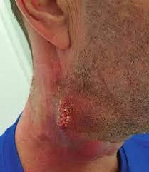
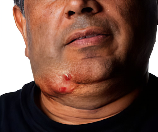
Bacillary Angiomatosis

Bacillary Angiomatosis: Understanding a Rare but Important Condition


Introduction

Bacillary angiomatosis (BA) is a rare vascular disease primarily caused by two species of bacteria: Bartonella henselae and Bartonella quintana. This condition primarily affects individuals with weakened immune systems and is often associated with HIV/AIDS. Despite its rarity, understanding bacillary angiomatosis is essential for timely diagnosis and treatment, especially in vulnerable populations.


ETIOLOGY

Causative Agents

The culprits behind bacillary angiomatosis are Bartonella henselae and Bartonella quintana. Bartonella henselae is commonly linked to cat scratches and bites, while Bartonella quintana is primarily transmitted through human interactions, particularly in overcrowded conditions.


Bartonella

Transmission Routes

Cat Scratches and Bites: The most common mode of transmission is through scratches or bites from infected cats. This is why bacillary angiomatosis is often seen in cat owners or those frequently around felines.

Human-to-Human Transmission: Although rare, Bartonella quintana can be transmitted between humans, primarily through close contact or in unsanitary conditions.


 Risk Factors

Individuals who are immunocompromised, particularly those with HIV/AIDS, are at a higher risk of developing bacillary angiomatosis. A compromised immune system allows the bacteria to proliferate, leading to the characteristic symptoms and lesions associated with the condition.


Pathophysiology

Bacillary angiomatosis starts when the bacteria invade the host's skin, usually following an injury or scratch. Once inside, they provoke an inflammatory response that leads to the formation of new blood vessels, a process known as angiogenesis. This abnormal growth results in the distinctive nodular and vascular lesions seen in affected individuals.


CLINICAL MANIFESTATIONS

Common Symptoms

Patients with bacillary angiomatosis may experience a range of symptoms, including:

  • Skin Lesions: The most noticeable signs are skin lesions that can appear as nodules or vascular lesions, often resembling tumors.
  • Fever and Malaise: General feelings of unwellness, including fever and fatigue, are also common.
  • Lymphadenopathy: Swelling of lymph nodes may occur, particularly near the site of infection.


Lesion Characteristics

  • Color and Texture: The lesions typically have a reddish or purplish color, with a soft, raised texture.
  • Location on the Body: While lesions can appear anywhere, they are most commonly found on the skin and subcutaneous tissues.


Complications if Left Untreated

If left untreated, bacillary angiomatosis can lead to serious complications, including systemic infections and complications related to the lesions themselves.


DIAGNOSIS

Clinical Evaluation

A thorough clinical evaluation, including a detailed medical history and physical examination, is the first step toward diagnosis.


Diagnostic Tests

  • Histopathological Examination: A biopsy of the lesions can provide definitive evidence of bacillary angiomatosis by revealing characteristic histological features.
  • Serological Tests: Blood tests can be performed to detect antibodies against Bartonella species.
  • Imaging Studies: If systemic involvement is suspected, imaging studies such as ultrasounds or CT scans may be utilized to assess internal organs.


BACILLARY ANGIOMATOSIS: TREATMENT AND PREVENTION

Bacillary angiomatosis is a bacterial infection that can be effectively treated with antibiotics, making early recognition crucial. Most treatment recommendations come from clinical observations and expert opinions, as there are no prospective studies or randomized clinical trials specific to this disease. For individuals with HIV, antiretroviral therapy is essential, though it can lead to complications such as immune reconstitution inflammatory syndrome and the Jarisch-Herxheimer reaction.


Surgical and Specialist Care 

In some cases, surgical treatments like cryotherapy or excision can help manage solitary skin lesions, but antibiotics remain key for addressing deeper infections. Specialists such as infectious disease experts or dermatologists may be consulted to provide the best care, especially for patients with complex or severe cases. Long-term monitoring is essential to track treatment effectiveness, particularly when internal organs are involved.


Preferred Antibiotic Therapy 

Erythromycin is the first-line treatment for bacillary angiomatosis due to its strong effectiveness. Tetracycline-based antibiotics, like doxycycline, are also effective alternatives for those who cannot tolerate erythromycin. In more severe cases, particularly in immunocompromised individuals, a combination of antibiotics such as doxycycline with rifampin may be required. Other antibiotics, like clarithromycin or azithromycin, have shown success in certain cases, but treatment failures have been reported with some options like ciprofloxacin or trimethoprim-sulfamethoxazole. The optimal length of therapy isn't well defined, but skin lesions usually respond within 4-7 days, with full resolution often taking up to a month. In patients with HIV, extended or lifelong treatment may be necessary if relapses occur.


PREVENTION STRATEGIES  

Preventing bacillary angiomatosis caused by Bartonella henselae involves minimizing exposure to cats, particularly through flea control. In cases involving Bartonella quintana, delousing procedures, like permethrin dusting powder, and maintaining clean clothing and bedding are recommended. Macrolide antibiotics used to prevent Mycobacterium avium-intracellulare in HIV patients also offer protection against bacillary angiomatosis.


CONCLUSION 

While bacillary angiomatosis is treatable with antibiotics, long-term monitoring and prevention, particularly in immunocompromised individuals, are key to managing and reducing recurrence of the disease. Consultation with medical specialists and careful use of antibiotics remain crucial for achieving the best outcomes.


ARCANOBACTERIUM HAEMOLYTICUM INFECTION (Hinglish)

ARCANOBACTERIUM HAEMOLYTICUM INFECTION

Arcanobacterium haemolyticum ek bacterium hai jo 1946 mein pehli baar identify kiya gaya tha. Yeh primarily pharyngitis aur skin rashes jaise infections ka kaaran banta hai, aur khaaskar US military personnel aur South Pacific ke indigenous communities mein paya gaya hai. Shuruaat mein ise Corynebacterium pyogenes ka subspecies mana gaya tha, lekin baad mein iske distinct characteristics ke kaaran ise nayi genus Arcanobacterium mein reclassify kiya gaya, jiska matlab hai "Is bacterium ko 'chhupa hua bacteria' kaha gaya. Vaqt ke sath, ise sepsis aur osteomyelitis jaise gambhir infections se bhi sambandhit kiya gaya hai."


SYMPTOMS AUR PRESENTATION

A. haemolyticum infection ka sabse common symptom sore throat hai, jo lagbhag sabhi patients (97-100%) mein report kiya jata hai. Dusre symptoms mein shamil hain:

  • Itchy skin (33%)
  • Nonproductive cough (33%)
  • Fever
  • Rash

Yeh rashes arms aur legs se shuru hote hain, lekin chehre, palms aur soles ko nahi chhute. Rash 48 ghante tak reh sakta hai, aur recovery ke dauran halki peeling dekhne ko milti hai. Ek review mein, 50% se zyada patients mein tonsillar exudates, fever, aur skin rash dekha gaya.


TRANSMISSION AUR RISK FACTORS

Ab tak koi confirmed risk factors nahi mile hain, lekin yeh speculate kiya gaya hai ki cows aur buffaloes jaise animals ke contact mein aane ya unpasteurized milk ke consumption se infection ka risk badh sakta hai. Bacterium European bison mein bhi paya gaya hai, lekin transmission ka tareeka ab tak unclear hai. Human-to-human transmission ka bhi shak hai, lekin proper hygiene, jaise handwashing, se iske spread ko roka ja sakta hai.


COMPLICATIONS

A. haemolyticum infection ke complications mein peritonsillar abscesses, sepsis, aur skin aur soft tissue infections shamil ho sakte hain. Severe sore throat aur fever ke cases mein hospitalization ki zarurat pad sakti hai jisme abscess drainage ya intravenous antibiotics ki madad li jaati hai.


DIAGNOSIS

A. haemolyticum ka diagnosis mushkil ho sakta hai, kyunki iski colonies slow-growing hoti hain aur routine throat cultures mein miss ho sakti hain. Specialized blood agar aur prolonged incubation (48 ghante tak) ki zarurat padti hai isse detect karne ke liye. Kuch cases mein, rashes se li gayi skin biopsies mein inflammation dikhayi deti hai, lekin bacteria khud nahi milta.


ARCANOBACTERIUM HAEMOLYTICUM INFECTION KA TREATMENT

A. haemolyticum ek significant lekin underrecognized bacterium hai jo mild pharyngitis se lekar severe invasive diseases, jaise sepsis ya osteomyelitis tak infections ka kaaran ban sakta hai. Jaldi pehchan aur proper treatment complications ka risk kam karne mein madad karte hain. Niche A. haemolyticum infections ke treatment ke bare mein comprehensive guide di gayi hai.


1. Pharyngitis aur Mild Infections

A. haemolyticum ke zyada infections pharyngitis ke roop mein hote hain, jisme tonsillitis, fever, aur kabhi-kabhi rash bhi dikhai dete hain. Halke cases khud theek ho sakte hain, lekin antibiotic treatment symptoms ki duration kam karne aur complications se bachne ke liye recommend kiya jata hai.

Antibiotic Therapy:

  • Penicillin: Penicillin aur related antibiotics jaise amoxicillin standard treatments hote hain, lekin kuch cases mein bacteria ke intracellular survive karne ke kaaran resistance dekhne ko milta hai.
  • Macrolides (e.g., erythromycin, azithromycin): Macrolides ko intracellular bacteria ke against effective mana jata hai, aur yeh A. haemolyticum infections ke treatment ke liye first choice hote hain. Azithromycin ko iski convenient dosing schedule ke liye khaaskar pasand kiya jata hai.
  • Clindamycin: Penicillin allergy wale patients ya severe infections wale patients ke liye ek viable alternative hai. Clindamycin achhi tissue penetration ke saath anaerobic bacteria ke against effective hota hai.

Dosage Recommendations:

  • Tab. Erythromycin: 250-500 mg har 6-12 ghante ke liye, 7-10 din tak.
  • Tab. Azithromycin: Pehle din 500 mg, uske baad 4 din tak 250 mg roz.
  • Tab. Clindamycin: 150-450 mg har 6-8 ghante, infection ki severity ke hisaab se.

Expected Outcomes: Antibiotic therapy se pharyngitis usually 3 din mein theek ho jata hai. Bina treatment ke symptoms 2 hafte tak chal sakte hain.


2. Severe ya Systemic Infections

Zyada severe cases, jaise sepsis ya deep tissue infections, mein jaldi intravenous antibiotics ka intervention zaroori hota hai.

Intravenous Antibiotics:

Beta-lactams (e.g., penicillin, cefuroxime, cefotaxime): Yeh antibiotics severe cases jaise sepsis ya osteomyelitis ke liye first-line treatments hain. Yeh bacterial cell wall ke construction mein interfere karke bacteria ko kamzor karte hain aur uske growth ko rok dete hain.

Vancomycin: Severe infections mein beta-lactams allergy wale patients ya resistance ke shak par use hota hai. Lekin vancomycin ke resistance ke cases bhi report kiye gaye hain, isliye sensitivity testing zaroori hai.

Antibiotic Sensitivity: A. haemolyticum typically sensitive hota hai:

  • Minocycline
  • Vancomycin
  • Beta-lactams

Antibiotic Resistance: Resistance dekhne ko mili hai:

  • Gentamicin
  • Fluoroquinolones (e.g., levofloxacin, ciprofloxacin)
  • Trimethoprim-sulfamethoxazole
  • Tetracyclines (30% strains mein resistance)

Treatment Duration: Severe infections ke liye intravenous therapy usually 2-6 hafton tak chalti hai, infection ki site aur severity ke hisaab se.


3. Complications ka Management

Kuch cases mein, untreated ya severe infections peritonsillar abscesses ya sepsis jaisi complications tak pahunch sakti hain.

  • Peritonsillar Abscess: Is condition mein surgical drainage ki zarurat padti hai, jiske baad antibiotics (penicillin, clindamycin, ya cefuroxime) 7-14 din tak diye jate hain.
  • Sepsis: Immediate intravenous antibiotics aur supportive care zaroori hota hai. Blood cultures sensitivity results ke hisaab se appropriate antibiotics select karne mein madad karte hain.


4. Adjunct Therapies

Supportive treatments antibiotic therapy ko complement kar sakti hain, khaaskar pharyngitis ya systemic infections ke cases mein.

  • Antipyretics aur Analgesics: Non-steroidal anti-inflammatory drugs (NSAIDs) jaise ibuprofen ya acetaminophen fever aur pain manage karne mein madad karte hain.
  • Antiseptic Lozenges: Amylmetacresol jaise antiseptics wale lozenges bacterial load kam karne aur symptoms ko soothe karne mein madad karte hain.


5. Hospital Admission aur Parenteral Therapy

  • Systemic ya severe infections wale patients ko hospitalization ki zarurat ho sakti hai, khaaskar agar wo swallow nahi kar pa rahe ho ya sepsis jaise complications ka risk ho.
  • Intravenous Antibiotics: Aise patients ko parenteral antibiotics ki zarurat hoti hai aur infection resolve hone tak close monitoring ki jati hai.


Monitoring aur Follow-Up

Patients ko antibiotics shuru karne ke 48-72 ghante ke andar reassess kiya jana chahiye taaki improvement ensure kiya ja sake. Severe cases mein, repeat blood cultures aur imaging required ho sakti hai recovery confirm karne ke liye.


PREVENTION

A. haemolyticum ke liye koi specific preventive measures recommended nahi hain, lekin achhi hygiene maintain karna, shared utensils avoid karna, aur proper food handling (khaaskar dairy products ke saath) infection transmission ke risk ko kam kar sakte hain.


CONCLUSION 

Early detection aur appropriate treatment critical hote hain Arcanobacterium haemolyticum infections ka sahi management aur complications ko prevent karne ke liye.

Arcanobacterium haemolyticum Infections

Arcanobacterium haemolyticum Infections

Arcanobacterium haemolyticum is a bacterium first identified in 1946, known for causing infections like pharyngitis and skin rashes, particularly in US military personnel and indigenous communities in the South Pacific. Initially thought to be a subspecies of Corynebacterium pyogenes, it was later reclassified into the new genus Arcanobacterium, meaning "secretive bacteria," due to its distinct characteristics. Over time, it has been linked to more serious infections, including sepsis and osteomyelitis.


Symptoms and Presentation

The most common symptom of A. haemolyticum infection is a sore throat, reported in almost all patients (97-100%). Other symptoms include:

  • Itchy skin (33%)
  • Nonproductive cough (33%)
  • Fever
  • Rash

The rash typically starts on the arms and legs, sparing the face, palms, and soles. The rash may last over 48 hours, with mild peeling during recovery. In a review of 191 cases, over 50% of patients experienced tonsillar exudates, fever, and skin rash.


Transmission and Risk Factors

Though no confirmed risk factors exist, it's been speculated that contact with animals like cows and buffaloes or consumption of unpasteurized milk may increase the risk of infection. The bacterium has also been found in European bison, although its transmission mechanism remains unclear. Human-to-human transmission is suspected, but proper hygiene, like handwashing, may help prevent its spread.


Complications

Complications from A. haemolyticum infection can range from peritonsillar abscesses, sepsis, and various skin and soft tissue infections. In some cases, severe sore throat and fever may lead to hospitalization for drainage of abscesses or intravenous antibiotics.


Diagnosis

Diagnosing A. haemolyticum can be tricky, as its colonies are slow-growing and may be missed in routine throat cultures. Specialized blood agar and prolonged incubation (up to 48 hours) are often needed to detect it. In some cases, skin biopsies from rashes show inflammation but do not contain the bacteria itself.


Treatment of Arcanobacterium haemolyticum Infection

Arcanobacterium haemolyticum is a significant yet underrecognized bacterium responsible for various infections, ranging from mild pharyngitis to severe invasive diseases such as sepsis or osteomyelitis. Early recognition and proper treatment are critical to minimizing the risk of complications. Below is a comprehensive guide to managing infections caused by A. haemolyticum.

1. Pharyngitis and Mild Infections

Most infections caused by A. haemolyticum manifest as pharyngitis, often presenting with tonsillitis, fever, and sometimes a rash. While mild cases may resolve spontaneously, antibiotic treatment is recommended to shorten symptom duration and avoid complications.

Antibiotic Therapy:

  • Penicillin: Although penicillin and related antibiotics like amoxicillin are standard treatments, some cases show resistance due to the bacterium’s ability to survive intracellularly.
  • Macrolides (e.g., erythromycin, azithromycin): Preferred for their effectiveness against intracellular bacteria, macrolides are often the first choice for treating A. haemolyticum infections. Azithromycin is especially favored for its convenient dosing schedule.
  • Clindamycin: This is a viable alternative for patients allergic to penicillin or those experiencing severe infections. Clindamycin has good tissue penetration and is effective against anaerobic bacteria.


Dosage Recommendations:

  • Tab. Erythromycin: 250-500 mg every 6-12 hours for 7-10 days.
  • Tab. Azithromycin: 500 mg on the first day, followed by 250 mg daily for 4 days.
  • Tab. Clindamycin: 150-450 mg every 6-8 hours, depending on severity.

Expected Outcomes: Antibiotic therapy generally resolves pharyngitis within 3 days. Without treatment, symptoms can longer for up to 2 weeks.

2. Severe or Systemic Infections

More severe cases, such as sepsis or deep tissue infections, demand prompt intervention with intravenous antibiotics.

Intravenous Antibiotics:

  • Beta-lactams (e.g., penicillin, cefuroxime, cefotaxime): These antibiotics are first-line treatments for severe cases like sepsis or osteomyelitis. They function by interfering with the construction of the bacterial cell wall, weakening the bacteria and preventing it from growing or surviving.
  • Vancomycin: Useful for severe infections in patients allergic to beta-lactams or when resistance is suspected. However, resistance to vancomycin has been reported, so sensitivity testing is essential.


Antibiotic Sensitivity: A. haemolyticum is typically sensitive to:

  • Minocycline
  • Vancomycin
  • Beta-lactams


Antibiotic Resistance: Resistance has been observed with:

  • Gentamicin
  • Fluoroquinolones (e.g., levofloxacin, ciprofloxacin)
  • Trimethoprim-sulfamethoxazole
  • Tetracyclines (30% resistance rate in tested strains)


Treatment Duration: For severe infections, intravenous therapy typically lasts 2-6 weeks, depending on the site and severity.


3. Management of Complications

In some cases, untreated or severe infections may lead to complications like peritonsillar abscesses or sepsis.

  • Peritonsillar Abscess: This condition requires surgical drainage followed by antibiotics (penicillin, clindamycin, or cefuroxime) for 7-14 days.
  • Sepsis: Immediate intravenous antibiotics and supportive care are crucial. Blood cultures guide the selection of appropriate antibiotics based on sensitivity results.


4. Adjunct Therapies

Supportive treatments can complement antibiotic therapy, particularly in cases of pharyngitis or systemic infections.

  • Antipyretics and Analgesics: Non-steroidal anti-inflammatory drugs (NSAIDs) like ibuprofen or acetaminophen help manage fever and pain.
  • Antiseptic Lozenges: Lozenges with antiseptics such as amylmetacresol can reduce bacterial load and soothe symptoms.


5. Hospital Admission and Parenteral Therapy

Hospitalization may be necessary for patients with systemic or severe infections, especially if they cannot swallow or are at risk of complications like sepsis.

Intravenous Antibiotics: These patients will require parenteral antibiotics and close monitoring to ensure the infection resolves.


 6. Monitoring and Follow-Up

Patients should be reassessed within 48-72 hours of starting antibiotics to ensure improvement. In severe cases, repeat blood cultures and imaging may be required to confirm recovery.


Prevention

While no specific preventive measures are recommended for A. haemolyticum, maintaining good hygiene, avoiding shared utensils, and proper food handling (particularly with dairy products) can help reduce transmission.

In conclusion, early detection and appropriate treatment are critical in managing Arcanobacterium haemolyticum infections and preventing complications.

Hand, Foot, and Mouth Disease in Children (HFMD)


Hand, Foot, and Mouth Disease in Children


Introduction 

Hand, foot, aur mouth disease (HFMD) ek common viral bimari hai jo mainly chhote bachchon ko affect karti hai. Yeh usually mild hoti hai, lekin kabhi-kabhi discomfort aur complications ka kaaran ban sakti hai. HFMD viruses ke enterovirus family ke karan hoti hai, jisme se coxsackievirus A16 aur enterovirus 71 sabse common hain.


Symptoms 

Virus ke exposure ke 3 se 7 din baad symptoms nazar aane lagte hain, jo include karte hain:

1. Fever: Pehla sign hota hai, jo usually mild se moderate hota hai.

2. Sore Throat: Gala mein dard ho sakta hai, aur nighalne mein bhi takleef ho sakti hai.

3. Mouth Sores: Dardnakh zakhm munh mein bante hain, jo zyadaatar jeebh, masoodon, aur andar ke gaalon par hote hain.

4. Rash: Haathon aur paon par ek rash bhi dikhayi de sakta hai. Yeh daag usually laal hote hain aur blister ban sakte hain.

5. Loss of Appetite: Munh ke sores ke wajah se bachche khana ya pani nhin lete hain.

6. Fatigue aur Irritability: General thakaan aur chidchidaapan hota hai, jo chhote bachchon mein zyada dikhai deta hai.


Transmission 

HFMD chhune se phailne waali bimari hai, aur yeh is tarah se spread hoti hai:

  • Direct Contact: Infected surfaces ya fluids (jaise saliva, blister ka fluid) ko touch karna.
  • Respiratory Droplets: Kisi bimaar shakhs ke khansi ya cheekhne se.
  • Feco-Oral Route: Virus stool mein bhi hota hai, to safai ki kami se yeh phail sakta hai.


Diagnosis 

Diagnosis usually in baaton par depend karta hai:

  • Clinical Evaluation: Doctor symptoms ko assess karte hain aur characteristic mouth sores aur rashes dekhte hain.
  • Recent Illness History: Schools ya daycare centers mein recent outbreaks ko note karna helpful ho sakta hai.
  • Kabhi-kabhi, laboratory tests ki zarurat ho sakti hai taaki virus ka type confirm ho sake.


Bacchon mein Hand, Foot, aur Mouth Disease (HFMD) ka ilaj 

HFMD ka koi specific antiviral treatment nahi hai, par symptoms ko manage karne aur supportive care dene par focus kiya jata hai. Yahan kuch key treatment strategies di gayi hain:

1. Symptom Relief 

Pain Relief - Over-the-Counter Medications:

  • Acetaminophen (Tylenol): Bukhar kam karne aur dard se rahat dene ke liye.
  • Ibuprofen (Advil, Motrin): Bukhar aur discomfort kam karne ka ek aur option.
  • Note: Bacchon ko aspirin dena avoid karein kyunki Reye's syndrome ka risk hota hai.

Mouth Sores:

  • Topical Anesthetics: Benzocaine wale gels ya liquids temporary dard ko numb kar sakte hain. Yeh sores par directly lagaye ja sakte hain.
  • Saltwater Rinses: Namak aur garam paani ka solution gala aur munh ke sores ko soothe karne mein madad karta hai (lekin chhote bachchon ke liye use karna mushkil ho sakta hai).

2. Hydration 

  • Fluid Intake Encourage Karein: Bacchon ko hydrated rakhna bahut zaruri hai, khaaskar jab unhe munh ke sores ke kaaran nighalne mein mushkil hoti hai.
  • Thande Fluids Offer Karein: Ice chips, popsicles, ya smoothies soothing ho sakte hain aur hydration mein madad karte hain.
  • Acidic ya Spicy Foods/Drinks Avoid Karein: Yeh munh ke sores ko irritate kar sakte hain.

3. Nutrition 

Soft Foods Encourage Karein: Aise foods khilayein jo soft aur easily nighalne wale ho, jaise:

  • Mashed potatoes
  • Applesauce
  • Yogurt
  • Oatmeal 
  • Hard ya Crunchy Foods Avoid Karein: Aise foods jo munh ke sores ko irritate kar sakte hain, avoid karein.

4. Fever Management

Temperature Monitor Karein: Bacchon ka temperature track karein aur zarurat padne par fever-reducing medications de.

Comfort Measures: Bacchon ko lightweight kapde pehnayein aur unke aas-paas ka environment thanda rakhein taaki fever ko manage kiya ja sake.

5. Rest aur Comfort

Rest Encourage Karein: Bacchon ko rest karne ke liye ek quiet aur comfortable environment dein.

Distraction Techniques: Aise activities jaise reading, quiet games, ya movies dekhna use karein taaki bacchon ko recovery ke dauraan busy rakha ja sake.

6. Follow-Up Care 

Symptoms par nazar rakhein: Bacchon ki tabiyat ko gahraai se observe karein. Agar lakshan bigadne lagein, pani ki kami, gambhir symptoms ya phir uncha bukhar dikhai de, toh turant chikitsak se salaah lein.


Healthcare Provider se Salah Len: Agar dehydration ya severe symptoms ka darr ho, khaaskar chhote bacchon ya jinhe already health issues hain, unme doctor se consult karna zaruri hai.


Prevention 

Preventive measures mein include hota hai:

  • Achhi Safai ki Adatein: Bathroom use karne ke baad aur khane se pehle sabun se haath dhona.
  • Close Contact Avoid Karein: HFMD wale bacchon ko school ya daycare se door rakhein jab tak wo contagious na ho (usually jab fever theek ho jaye aur munh ke sores heal ho jaayein).
  • Surfaces Disinfect Karein: Toys aur frequently touched surfaces ko clean karke virus ke spread ko kam kiya ja sakta hai.


Complications 

HFMD usually mild hota hai, lekin kabhi-kabhi complications bhi ho sakte hain, jaise:

  • Dehydration: Munh ke sores ke kaaran nighalne mein dikkat ke karan.
  • Viral Meningitis: Kabhi-kabhi virus se serious neurological conditions ho sakti hain.
  • Myocarditis ya Encephalitis: Yeh serious complications rare hote hain, lekin enterovirus infections mein ho sakte hain.


Prognosis 

HFMD usually 7 se 10 din mein bina medical intervention ke theek ho jata hai. Zyadaatar bacche bina koi long-term health issues ke fully recover karte hain.


Conclusion 

Hand, foot, aur mouth disease ek common, generally mild viral infection hai bacchon mein. Symptoms, transmission, aur preventive measures ka dhyaan rakhke outbreaks ko effectively manage kiya ja sakta hai, aur illness ke dauraan bacchon ko comfortable rakha ja sakta hai. Agar severe symptoms ya dehydration ka sign dikhe, toh turant medical attention leni chahiye.


Hand, Foot, and Mouth Disease in Children


Hand, Foot, and Mouth Disease in Children


Introduction

Hand, foot, and mouth disease (HFMD) is a common viral illness primarily affecting young children. It is usually mild but can cause discomfort and occasionally lead to complications. HFMD is caused by viruses from the enterovirus family, with coxsackievirus A16 and enterovirus 71 being the most common culprits.


Symptoms:

Symptoms typically appear 3 to 7 days after exposure to the virus and may include:

1. Fever: Often the first sign, usually mild to moderate.

2. Sore Throat: Can be accompanied by pain when swallowing.

3. Mouth Sores: Painful lesions develop in the mouth, often on the tongue, gums, and inner cheeks.

4. Rash: A rash may appear on the hands and feet. The spots are typically red and may blister.

5. Loss of Appetite: Due to mouth sores, children may refuse to eat or drink.

6. Fatigue and Irritability: General malaise is common, leading to fussiness in younger children.


Transmission

HFMD is contagious and spreads through:

Direct Contact: Touching infected surfaces or secretions (saliva, fluid from blisters).

Respiratory Droplets: Coughing or sneezing from an infected person.

Feco-Oral Route: The virus can be present in stool, so inadequate hygiene practices can facilitate spread.


Diagnosis

Diagnosis is generally based on:

  • Clinical Evaluation: Healthcare providers assess symptoms and look for characteristic mouth sores and rashes.
  • History of Recent Illness: Noting any recent outbreaks in schools or daycare centers can aid diagnosis.
  • In rare cases, laboratory tests may be performed to confirm the virus type.


Treatment of Hand, Foot, and Mouth Disease (HFMD) in Children

Although there is no specific antiviral treatment for HFMD, management focuses on alleviating symptoms and providing supportive care. Here are the key treatment strategies:

 1. Symptom Relief

Pain Relief - OvertheCounter Medications:

  • Acetaminophen (Tylenol): Helps reduce fever and relieve pain.
  • Ibuprofen (Advil, Motrin): Another option for reducing fever and discomfort.
  • Note: Avoid aspirin in children due to the risk of Reye's syndrome.

Mouth Sores:

  • Topical Anesthetics: Gels or liquids containing benzocaine may numb the pain temporarily. These can be applied directly to the sores.
  • Saltwater Rinses: A solution of salt and warm water can help soothe the throat and mouth sores (but may be difficult for young children to use).

 2. Hydration

  • Encourage Fluid Intake: It's crucial to keep children hydrated, especially if they have difficulty swallowing due to mouth sores.
  • Offer Cool or Cold Fluids: Ice chips, popsicles, or smoothies can be soothing and help with hydration.
  • Avoid Acidic or Spicy Foods/Drinks: These can irritate the mouth sores further.

 3. Nutrition

Soft Foods: Encourage the consumption of soft, bland foods that are easy to swallow, such as:

  • Mashed potatoes
  • Applesauce
  • Yogurt
  • Oatmeal
  •  Avoid Hard or Crunchy Foods: Foods that may cause irritation to mouth sores should be avoided.

4. Fever Management

  • Monitor Temperature: Keep track of the child's temperature, and administer feverreducing medications as needed.
  • Comfort Measures: Dress the child in lightweight clothing and keep their environment cool to help manage fever.

 5. Rest and Comfort

  • Encourage Rest: Provide a quiet, comfortable environment for the child to rest and recover.
  • Distraction Techniques: Use activities like reading, quiet games, or watching movies to keep the child occupied while they recover.

 6. FollowUp Care

  • Monitor Symptoms: Keep an eye on the child's condition. If symptoms worsen or if there are signs of complications (such as difficulty swallowing, severe dehydration, or a high fever persisting beyond a few days), seek medical attention.
  • Consult a Healthcare Provider: If there are concerns about dehydration or severe symptoms, especially in very young children or those with underlying health conditions.


Prevention

Preventive measures include:

  • Good Hygiene Practices: Regular handwashing with soap and water, especially after using the restroom and before eating.
  • Avoiding Close Contact: Keeping children with HFMD away from school or daycare until they are no longer contagious (usually when fever resolves and mouth sores heal).
  • Disinfecting Surfaces: Cleaning toys and frequently touched surfaces can reduce the spread of the virus.


Complications

While HFMD is usually mild, complications can arise, including:

  • Dehydration: Resulting from difficulties in swallowing due to mouth sores.
  • Viral Meningitis: Rarely, the virus can lead to more severe neurological conditions.
  • Myocarditis or Encephalitis: These serious complications are very rare but can occur with enterovirus infections.


Prognosis

HFMD typically resolves within 7 to 10 days without medical intervention. Most children recover completely with no longterm health issues.


Conclusion

Hand, foot, and mouth disease is a common, typically mild viral infection in children. Awareness of symptoms, transmission, and preventive measures can help manage outbreaks effectively and keep children comfortable during illness. If a child shows severe symptoms or signs of dehydration, medical attention should be sought.

ACTINOMYCOSIS (Hinglish)

 ACTINOMYCOSIS


Introduction:   

Actinomycosis ek chronic bacterial infection hai jo 'Actinomyces' bacteria se hota hai. Ye bacteria filamental, gram-positive hote hain, jo aam taur pe mouth, digestive system, aur female genital tract mein paye jaate hain. Lekin jab mucous membrane damage hoti hai, jaise dental surgery, abdominal injury, ya IUD use karne ke baad, to infection shuru ho sakta hai.


Actinomycosis kaise develop hota hai:   

Ye infection dheere dheere tissues mein failta hai aur abscesses aur sinus tracts banata hai. Kabhi kabhi is infection ke discharge mein sulfur granules (chhoti yellow particles) bhi dekhe ja sakte hain. Ye infection alag-alag jagah affect kar sakta hai jaise face (cervicofacial), chest (thoracic), ya abdomen aur pelvis. Isko diagnose karna mushkil hota hai kyunki ye tumors ya tuberculosis jaisi bimariyon jaise lagta hai.


Common types:   

1.  Cervicofacial Actinomycosis (Lumpy Jaw): Ye sabse common type hai aur aksar poor dental hygiene ya oral surgery se judta hai. Jaw mein swelling hoti hai, fistulas ban jaate hain, aur pus discharge hota hai jisme sulfur granules hoti hain.

2.  Thoracic Actinomycosis: Ye lungs ko affect karta hai, aksar bacteria inhale karne ke baad. Chest pain, cough, aur kabhi kabhi chest wall pe sinus tracts bhi ban jate hain.

3.  Abdominal aur Pelvic Actinomycosis: Surgery ya abdominal injury ke baad hota hai ya phir women mein IUD use ke wajah se hota hai. Abdominal pain, bowel habits mein changes, aur mass ki tarah feel hota hai, jise galat tareeke se cancer samajh liya jaata hai.


Symptoms:

  1. Cervicofacial Actinomycosis:  Jaw mein swelling, skin discoloration, sinus discharge, aur chewing mein dikkat.
  2. Thoracic Actinomycosis:  Cough, chest pain, shortness of breath, weight loss, aur fever.
  3. Abdominal Actinomycosis:  Abdominal pain, nausea, weight loss, aur abdomen mein mass ka sensation.
  4. Pelvic Actinomycosis:  Lower abdominal pain, vaginal bleeding ya discharge.


Diagnosis:   

Iska diagnosis karna thoda challenging hota hai. Abscesses ya sinus tract se tissue samples lene padte hain aur sulfur granules discharge mein milte hain to infection ka confirmation hota hai. Imaging tests jaise CT scan mass-like infection dikhate hain jo cancer jaise lag sakte hain. Laboratory tests anemia, elevated inflammatory markers show karte hain aur 'Actinomyces' ko cultures mein detect kar sakte  hain.


Treatment for Actinomycosis:

Is infection ka treatment antibiotics ke zariye kiya jaata hai.  Penicillin G  sabse zyada effective hoti hai. Treatment mein high doses antibiotics ka istamaal kiya jaata hai jo 6 mahine se lekar 1 saal tak chal sakta hai. Severe cases mein antibiotics ke saath surgery bhi zaroori ho sakti hai.


Antibiotic Treatment:

1. Penicillin G (Pfizerpen, Bicillin): Severe cases mein pehle intravenous (IV) Penicillin G diya jaata hai. Phir, oral antibiotics jaise Penicillin VK pe switch kiya jaata hai jab tak treatment complete na ho. Treatment 6 se 12 mahine tak chal sakta hai, ye infection ki severity aur response pe depend karta hai.

2.  Alternative antibiotics: Agar kisi ko penicillin se allergy ho, to doosre antibiotics ka istamaal kiya jaata hai jaise:

  • Doxycycline:  Ye bacterial protein synthesis ko rokta hai. Penicillin-allergic patients ke liye use hota hai, lekin pregnant women ke liye avoid kiya jaata hai.
  • Clindamycin (Cleocin):  Penicillin-allergic patients ke liye doosra option. Ye bacterial protein synthesis block karta hai, lekin companion bacteria ko cover nahi kar pata.
  • Amoxicillin/Clavulanic acid (Augmentin):  Mild to moderately severe cases mein use hota hai.
  • Ceftriaxone (Rocephin):  Moderate to severe infections mein use hota hai, jaise cervicofacial aur thoracic actinomycosis.
  • Imipenem/Cilastatin (Primaxin):  Severe abdominal ya pelvic infections ke liye kaafi effective hota hai.


Surgical Treatment:   

Jahan antibiotics se kaam nahi hota, wahan surgery zaroori ho sakti hai. Surgery ke zariye bade abscesses drain kiye jaate hain, infected tissue remove kiya jaata hai, aur kuch complications jaise fibrotic lesions ko treat kiya jaata hai. Kabhi kabhi abdominal actinomycosis mein patients ki emergency surgery ki zaroorat hoti hai agar peritonitis ya bowel perforation ho jaaye.


Monitoring During Treatment:   

Kyuki treatment kaafi lambe time tak chal sakta hai, regular follow-up zaroori hota hai jaise CT scan ya MRI ke zariye infection ka response monitor karna. Blood tests bhi karaye jaate hain recovery aur side effects dekhne ke liye.


Prevention:   

Achhi oral hygiene aur regular dental care important hai, khaaskar oral surgery ke baad. Aur IUD use karne wali women ko pelvic actinomycosis ke risk ko dhyaan mein rakhna chahiye.


Complications:   

Agar untreated chhoda gaya, to actinomycosis bone infections, brain abscesses, aur endocarditis (heart infection) jaise serious complications tak le ja sakta hai. Lekin early symptoms ko pehchaankar aur prolonged antibiotics course follow karke zyada tar patients achhe se recover karte hain.


ACTINOMYCOSIS


ACTINOMYCOSIS

Introduction

Actinomycosis is a chronic bacterial infection caused by ‘Actinomyces’ species, which are filamentous, gram-positive bacteria. These bacteria are normally present in the mouth, digestive system, and female genital tract, but they can cause infection when the mucous membranes are broken, typically after dental surgery, abdominal injury, or in women using intrauterine devices (IUDs).


How Actinomycosis Develops:

Actinomycosis spreads slowly through tissues, forming abscesses, sinus tracts, and sometimes discharging sulfur granules (tiny yellow particles).

The infection can involve various areas, such as the face (cervicofacial), chest (thoracic), or abdomen and pelvis. It can be difficult to diagnose since it mimics other diseases, including tumors and tuberculosis.


Common Types:

1.  Cervicofacial Actinomycosis (Lumpy Jaw) :

  • Most common, linked to poor dental hygiene or oral surgery.
  • It leads to soft swelling in the jaw, fistulas, and pus discharge containing sulfur granules.

2.  Thoracic Actinomycosis :

  • Affects the lungs, often after aspiration (inhaling bacteria from the mouth).
  • Presents as a lung mass, with chest pain, cough, and sometimes sinus tracts on the chest wall.

3.  Abdominal and Pelvic Actinomycosis :

  • Typically arises after surgery or trauma to the abdomen or from IUD use in women.
  • May cause abdominal pain, changes in bowel habits, and a growing mass, often misdiagnosed as cancer.


Symptoms:

  • Cervicofacial Actinomycosis : Jaw swelling, skin discoloration, sinus drainage, difficulty chewing.
  • Thoracic Actinomycosis : Cough, chest pain, shortness of breath, weight loss, and fever.
  • Abdominal Actinomycosis : Vague abdominal pain, nausea, weight loss, and a sensation of a mass in the abdomen.
  • Pelvic Actinomycosis : Lower abdominal pain, abnormal vaginal bleeding or discharge.


Diagnosis:

  • Diagnosing actinomycosis can be challenging, often requiring tissue samples from abscesses or sinus tracts. Sulfur granules in discharge are a key indicator.
  • Imaging tests (like CT scans) may show mass-like infections, often confused with cancer.
  • Laboratory tests may reveal anemia, elevated inflammatory markers, and the presence of ‘Actinomyces’ in cultures.


Treatment for Actinomycosis

Actinomycosis is primarily treated with antibiotics, and Penicillin G is the preferred drug. The treatment usually involves high doses of antibiotics for a prolonged period, ranging from 6 months to 1 year. This extended duration is necessary because actinomycosis can be slow to respond to therapy, and it may require a combination of antibiotic and surgical interventions depending on the severity of the disease.

Antibiotic Treatment:

1.  Penicillin G  (Pfizerpen, Bicillin) is the  drug of choice (DOC) :

Initial phase: Treatment typically starts with intravenous (IV) Penicillin G, especially in severe cases. A central catheter (PICC line) may be used to administer antibiotics over weeks to months.

Oral transition: After the initial IV treatment, patients are switched to oral antibiotics, such as Penicillin VK, for the remaining course of therapy.

Duration: The total treatment duration can range from 6 to 12 months, depending on how the infection responds to therapy and the location of the disease.

2.  Alternative antibiotics: In patients allergic to penicillin or where penicillin may not be effective, the following alternatives can be used:

  • Doxycycline: A tetracycline antibiotic that inhibits bacterial protein synthesis. It is commonly used for patients who are allergic to penicillin but should be avoided in pregnant women.
  • Clindamycin  (Cleocin): Another option for penicillin-allergic patients. Clindamycin blocks bacterial protein synthesis, but it may not cover all companion bacteria that often accompany actinomyces infection.
  • Amoxicillin/Clavulanic acid  (Augmentin): This combination can be used in  mild to moderately severe cases. It covers both the actinomyces bacteria and their companion bacteria.
  • Ceftriaxone  (Rocephin): A third-generation cephalosporin, useful in  moderate to severe infections, especially cervicofacial and thoracic actinomycosis.
  • Imipenem/Cilastatin  (Primaxin): Often used in  severe abdominal or pelvic infections , this combination antibiotic can target both actinomyces and other bacteria resistant to penicillin.


Surgical Treatment:

In addition to antibiotics, surgery may be necessary in certain cases. Surgery is typically reserved for:

  • Draining large abscesses or sinus tracts.
  • Removing infected tissue that does not respond well to antibiotics.
  • Treating complications such as obstructions caused by fibrotic lesions or infections compressing vital structures (e.g., ureters)

For example, patients with abdominal actinomycosis  may require emergency surgery for conditions like peritonitis or bowel perforation. Similarly, surgery may be needed to decompress infections in closed spaces, such as within the thoracic cavity or near the brain.


Monitoring During Treatment:

Since treatment can last several months, regular follow-up with imaging studies like CT scans or MRI is essential to monitor the response to therapy. Blood tests  may also help track the patient’s recovery and watch for side effects. In some cases, especially when using intravenous antibiotics, a PICC line may be inserted to facilitate prolonged antibiotic administration at home.

Other Considerations :

If Penicillin G therapy fails, it could be due to companion bacteria that resist penicillin. In such cases, the antibiotic regimen might need to be broadened to include drugs that target these other bacteria.

Companion bacteria (such as Peptostreptococcus, Fusobacterium, or Prevotella species) often contribute to the infection and may require a tailored approach with additional antibiotics.

In summary, the treatment of actinomycosis is centered around prolonged, high-dose antibiotic therapy, often starting with IV penicillin and transitioning to oral antibiotics for several months. In more serious or complex cases, surgery might be needed to remove infected tissue or drain abscesses that don’t heal with antibiotics alone. Close monitoring with imaging and adjustments in the antibiotic regimen may be required to ensure the infection is fully controlled.


Prevention:

  • Good oral hygiene and regular dental care are essential, especially after oral surgery.
  • Women using IUDs should be aware of the increased risk of pelvic actinomycosis.


Complications:

If untreated, actinomycosis can lead to serious complications, such as bone infections, brain abscesses, and endocarditis (heart infection).

By recognizing early symptoms and following through with prolonged antibiotic treatment, most patients recover well.

Acrodermatitis Chronica Atrophicans (Hinglish)


 Acrodermatitis Chronica Atrophicans (ACA)


Introduction:

Acrodermatitis chronica atrophicans (ACA) European Lyme borreliosis ka late ya third stage hai, jo 'Borrelia afzelii' ke infection se hota hai. Is condition ko pehli baar 1883 mein identify kiya gaya aur 1902 mein describe kiya gaya. ACA ek progressive skin condition hai jisme fibrosis aur tissue atrophy hoti hai, jismein tissue paper jaise lagte hain.


Pathophysiology aur Etiology of Acrodermatitis Chronica Atrophicans (ACA)

Causative Agents:

ACA ka primary cause Borrelia afzelii hota hai, lekin Borrelia garinii (Borrelia burgdorferi sensu lato complex ka dusra genospecies) bhi implicated hai. Ye spirochetes humans mein tick ke kaatne se transfer hote hain aur Lyme borreliosis ka cause bante hain.


aca
Borrelia afzelii


 Pathophysiology:

Lyme disease ke doosre forms ke opposite, ACA khud se thik nahi hoti, jo ek unique pathogenic mechanism ko dikhata hai. Iska exact process abhi tak puri tarah se samjha nahi gaya, lekin kuch factors Borrelia ke skin mein persist karne ke liye zimmedar hain:

1.  Spirochete Persistence:

Complement system ke resistance: Bacteria immune response se bache rehte hain.

Immune evasion: Spirochetes immunologically protected sites jaise endothelial cells aur fibroblasts mein chhupe rehte hain.

Antigenic variation: Pathogen apne surface antigens ko badal leta hai, jisse immune system effective response nahi de pata.

2.  Immune Dysfunction:

  • Inadequate antibody response: ACA patients ke antibodies ka spectrum narrow hota hai aur protective antibodies nahi bante.
  • MHC Class II molecules ka downregulation: Langerhans cells (jo skin ke immune cells hote hain) mein in molecules ki kami T-cells activation ko impair karti hai.
  • Cytokine expression: Restricted cytokine profile jaise interferon-gamma ka absence, immune system ko infection se effectively fight karne se rokta hai.

3.  Autoimmune Considerations:  Cross-reactive antibodies tissue damage mein yogdan karte hain, lekin autoimmunity ka role abhi clear nahi hai. Chronicity ek aise immune response se judi ho sakti hai jo inadvertently host tissues ko harm karta hai.


Atrophic Skin Changes ka Pathogenesis:

Exact mechanism jo ACA ke characteristic skin atrophy ka cause banta hai wo samjha nahi gaya hai, lekin yeh mana jata hai ki periarticular sites low skin temperatures ya oxygenation ki wajah se Borrelia spirochetes ke liye favorable environment provide karate hain.


Clinical Presentation:

ACA aksar extremities pe aati hai, khaas taur pe extensor surfaces par, aur yeh do distinct phases mein hoti hai:

1.  Inflammatory Phase:

  • Bluish-red skin discoloration aur swelling.
  • Soft, painless, poorly demarcated plaques jo milkar ek ho sakti hain.
  • Condition distal extremities se start hokar proximally spread hoti hai.


2.  Atrophic Phase:

  • Mahino ya saalon baad hoti hai, jisme dark red ya brown discoloration hoti hai.
  • Skin thin, translucent, aur wrinkled ho jati hai, cigarette paper jaisi dikhai deti hai.
  • Prominent blood vessels, hair loss, aur sebaceous ya sweat glands ka absence hota hai.


Associated Symptoms:

Fibrotic nodules elbows, knees, aur adjacent joints pe form hote hain. Skin ulcerate kar sakti hai, aur malignant lesions develop ho sakte hain. Fingers aur toes mein deformities bhi ho sakti hain agar untreated chhod diya jaye.

Systemic Involvement:

Neurological involvement common hai, jisme sensory polyneuropathy symptoms jaise pain, paresthesia, aur paresis (partial paralysis) shamil hote hain.


Diagnosis:

Detailed clinical history, epidemiologic data, histopathology aur serologic testing ke through diagnosis kiya jata hai. ELISA, indirect immunofluorescence, aur Western blot assays commonly use kiye jaate hain Borrelia burgdorferi antibodies ko detect karne ke liye.


Management of Acrodermatitis Chronica Atrophicans (ACA)


Approach to Treatment:

1.  Antibiotic therapy:

  • Oral antibiotics pehla-line treatment hoti hai jaise:
  • Doxycycline (100 mg do baar roz, 3-4 hafte ke liye).
  • Amoxicillin (500 mg teen baar roz, 3-4 hafte ke liye).

2.  Advanced ya Systemic Lyme disease:  Intravenous antibiotics jaise Ceftriaxone (2 g daily for 21-28 days) prefer kiye jaate hain.

3.  Complications ka Management: Fibrotic nodules aur joint deformities agar untreated rehte hain toh physiotherapy helpful ho sakti hai.

4.  Consultations for Systemic Involvement:  Neurologic, rheumatologic, ya cardiac signs ke liye specialists ki zarurat hoti hai.

5.  Coinfections aur Differential Diagnosis: ACA patients mein babesiosis ya ehrlichiosis jaise coinfections bhi ho sakte hain jo tick vector se transmitted hote hain.


Long-Term Monitoring:  

Patients ko follow-up mein rakha jata hai disease ke progression aur treatment ke effectiveness ko monitor karne ke liye. Atrophic phase mein skin changes persist kar sakte hain.


Patient Education:  

Patients ko batana chahiye ki ACA ka improvement weeks ya months le sakta hai. Lyme borreliosis ka early diagnosis aur treatment bahut zaroori hai, khaaskar endemic areas mein.


Prognosis:  

Early treatment se complete recovery possible hai, lekin atrophic phase ke symptoms partial reverse ho sakte hain.


Conclusion:  

ACA ek rare but significant manifestation hai Lyme borreliosis ka, jisme timely diagnosis aur appropriate treatment bahut zaroori hai taaki long-term complications se bacha ja sake. Antibiotic therapy effective hai, lekin early intervention irreversible damage se bacha sakti hai.

Acrodermatitis Chronica Atrophicans (ACA)


Acrodermatitis Chronica Atrophicans (ACA)


Introduction:

Acrodermatitis chronica atrophicans (ACA) is the late or third stage of European Lyme borreliosis, caused by an ongoing infection with ‘Borrelia afzelii’ . Initially identified in 1883 and described in 1902, ACA is a progressive skin condition characterized by fibrosis and tissue atrophy, resembling tissue paper in its appearance.


ACA


Pathophysiology and Etiology of Acrodermatitis Chronica Atrophicans (ACA)


Causative Agents:

Acrodermatitis chronica atrophicans (ACA) is primarily caused by Borrelia afzelii , though  Borrelia garinii , another genospecies from the  Borrelia burgdorferi sensu lato  complex, has also been implicated. These spirochetes are transmitted to humans via tick bites and are the underlying cause of Lyme borreliosis.


Borrelia afzelii
Borrelia afzelii


Pathophysiology:

Unlike other forms of Lyme disease, ACA does not resolve spontaneously, suggesting a unique pathogenic mechanism. The exact process through which ACA develops is not fully understood, but several factors contribute to the persistence of  Borrelia  in the skin:

1.  Spirochete Persistence:  Despite strong immune responses, the Borrelia spirochetes persist in the skin. This persistence might be due to:

  • Resistance to the complement system:  This allows the bacteria to evade destruction by immune responses.
  • Immune evasion:  The spirochetes can escape into immunologically protected sites, such as endothelial cells and fibroblasts, making it difficult for the immune system to target and eliminate them.
  • Antigenic variation:  The pathogen may change its surface antigens, causing the immune system to fail in mounting an effective or appropriate response.

2.  Immune Dysfunction:

  • Inadequate antibody response:  Patients with ACA exhibit a narrow spectrum of antibodies and lack sufficient protective antibodies. The immune system’s response, particularly cellular immunity, may be weakened.
  • Downregulation of MHC Class II molecules:  Reduced expression of these molecules on Langerhans cells (immune cells in the skin) impairs the activation of T-cells, crucial for an effective immune response.
  • Cytokine expression:  ACA is associated with a restricted cytokine profile, including the absence of interferon-gamma, which may prevent the immune system from clearing the infection effectively.

3.  Autoimmune Considerations:  Cross-reactive antibodies may contribute to tissue damage, though the role of autoimmunity in ACA remains unclear. The chronicity of the condition may involve an immune response that inadvertently harms the host tissues.


Pathogenesis of Atrophic Skin Changes:

  • The exact mechanism behind the characteristic skin atrophy in ACA is not well understood. However, it is hypothesized that:
  • Periarticular sites  may be particularly affected due to lower skin temperatures or reduced oxygenation in these areas, which could create a favorable environment for the persistence of  Borrelia  spirochetes.
  • Influence of Untreated Lyme Borreliosis: Failure to treat early-stage Lyme borreliosis adequately allows the infection to progress to ACA. This underscores the importance of early detection and treatment to prevent the chronic, progressive skin damage associated with ACA.


Clinical Presentation:

ACA usually manifests on the extremities, especially on the extensor surfaces, and follows two distinct phases:

1.  Inflammatory Phase:

  • Involves bluish-red discoloration and swelling of the skin.
  • Patients may develop soft, painless, poorly demarcated plaques that can coalesce.
  • The condition typically starts on the distal extremities and spreads proximally.


2.  Atrophic Phase:

  • Occurs months or years later, characterized by dark red or brown discoloration.
  • The skin becomes thin, translucent, and wrinkled, resembling cigarette paper.
  • Skin atrophy is associated with prominent blood vessels, loss of hair, and absence of sebaceous or sweat glands.
  • Telangiectasia and focal hyperpigmentation may be present.


aca


aca


Associated Symptoms:

  • Fibrotic nodules can form, usually on the extensor surfaces of the elbows, knees, and adjacent joints.
  • Skin may ulcerate, and malignant lesions can develop.
  • ACA can also cause deformities in the fingers and toes if untreated.

Systemic Involvement:

Neurological involvement is common in ACA, often presenting as sensory polyneuropathy. Symptoms such as pain, paresthesia, and even paresis (partial paralysis) can occur, and patients may exhibit abnormal vibratory thresholds. While cerebrospinal fluid abnormalities are rare, localized neuropathy often correlates with visible ACA lesions on the extremities.

Diagnosis:

Diagnosis relies on a detailed clinical history, epidemiologic data, and confirmation via histopathology and serologic testing. ELISA, indirect immunofluorescence, and Western blot assays are commonly used to detect Borrelia burgdorferi antibodies.


Management of Acrodermatitis Chronica Atrophicans (ACA)

The treatment of ACA, the late-stage manifestation of Lyme borreliosis, focuses on eradicating the infection and addressing any complications related to the disease. The choice of therapy depends on the presence of other signs of Lyme disease, the stage of ACA, and the results of serologic testing.

Approach to Treatment:

1.  Antibiotic therapy:

  • Oral antibiotics  are typically the first-line treatment in ACA, especially in the absence of systemic Lyme disease manifestations. Common options include:
  • Doxycycline  (100 mg twice daily for 3-4 weeks).
  • Amoxicillin  (500 mg three times daily for 3-4 weeks), especially in children or those allergic to doxycycline.
  • For patients with more advanced or systemic Lyme disease or those with high levels of antibodies, intravenous antibiotics  are preferred. These include:
  • Ceftriaxone  (2 g daily for 21-28 days).
  • Cefotaxime  or  Penicillin G  are also used in some cases.

Studies suggest that many patients, even in the chronic stage of ACA, can benefit from antibiotic treatment. In some patients, symptoms completely resolve after a 3-4 week course of IV ceftriaxone.

2.  Early vs. Late Stage Treatment:

In the  early inflammatory stage  of ACA, prompt antibiotic treatment can lead to full recovery and prevent further skin and tissue damage.

In the  chronic atrophic phase , while antibiotics can stop the progression of the disease, many of the skin changes (like atrophy and fibrosis) may be  irreversible . Patients should be informed that improvement may be partial.

3.  Management of Complications:  

Fibrotic nodules  and  joint deformities  can occur if ACA progresses untreated. In these cases, physical therapy may help improve joint mobility and prevent further deformity.

Ulcerations  on atrophic skin are common and can be challenging to treat. Proper wound care is essential, and in some cases, dermatologic consultation may be required for managing these lesions.

4.  Consultations for Systemic Involvement:

If extracutaneous signs of Lyme disease are present (neurologic, rheumatologic, or cardiac), appropriate consultations with specialists (neurologist, rheumatologist, cardiologist) should be made.

Neurologic symptoms , such as sensory polyneuropathy, may require further evaluation with neurophysiologic tests and long-term monitoring.

5.  Coinfections and Differential Diagnosis:

ACA patients may also have  co-infections  like babesiosis or ehrlichiosis, which are transmitted by the same tick vector. In regions where these infections are prevalent, testing for these conditions should be considered, especially if symptoms persist despite appropriate Lyme treatment.

Coinfections require additional treatments, for example,  atovaquone  and  azithromycin  for babesiosis or  doxycycline  for ehrlichiosis.


6.  Vaccination:

While the  US FDA  approved a vaccine for Lyme disease in 1999, it is no longer available. A new  recombined vaccine  targeting  B. afzelii  and  B. garinii , the prevalent strains in Europe, is being developed but is not yet in widespread use.


Long-Term Monitoring:

Patients should be followed up to monitor the progression of the disease and the effectiveness of treatment. In the atrophic phase, even though antibiotics may halt progression, skin changes may persist.

Regular skin checks  are recommended to monitor for any complications such as ulcers or skin cancers, as there is a small but present risk of malignancy in long-standing ACA cases.


Patient Education:

Patients should be informed that ACA may take several weeks or months to improve after the start of treatment. Improvement in symptoms may be gradual.

Early diagnosis and treatment of  Lyme borreliosis  are crucial to prevent the development of ACA, especially in endemic regions.

Prognosis:

Early treatment can result in complete resolution, though the atrophic phase symptoms may only partially reverse. Patients in the inflammatory phase should expect gradual symptom relief over several weeks or months following treatment.


Conclusion:

ACA is a rare but significant manifestation of Lyme borreliosis, requiring timely diagnosis and appropriate treatment to prevent long-term complications. While antibiotic therapy is effective, early intervention is key to preventing irreversible skin and nerve damage.

My Blog List